Description
Human USAG-1 (SOSTDC1) Protein, Fc Tag
Recombinant Human USAG-1 (SOSTDC1) Protein with C-terminal human IgG1 Fc tag, expressed in HEK293 cells and supplied lyophilized for research use.
Structure:

Human USAG-1 (SOSTDC1) Protein, Fc Tag
Highest Development Stage:
Preclinical
Product Overview
Synonym: Uterine sensitization-associated gene 1 protein
Also known as: SOSTDC1 (Sclerostin domain-containing protein 1), Ectodin
Human USAG-1 Protein, Fc Tag is a recombinant glycoprotein corresponding to amino acids Phe24–Ser206 (Accession # Q6X4U4-1). The protein is fused to a human IgG1 Fc tag at the C-terminus to enhance stability, solubility, and dimerization.
Produced in HEK293 (human embryonic kidney 293) cells, ensuring mammalian post-translational modifications and appropriate glycosylation patterns.
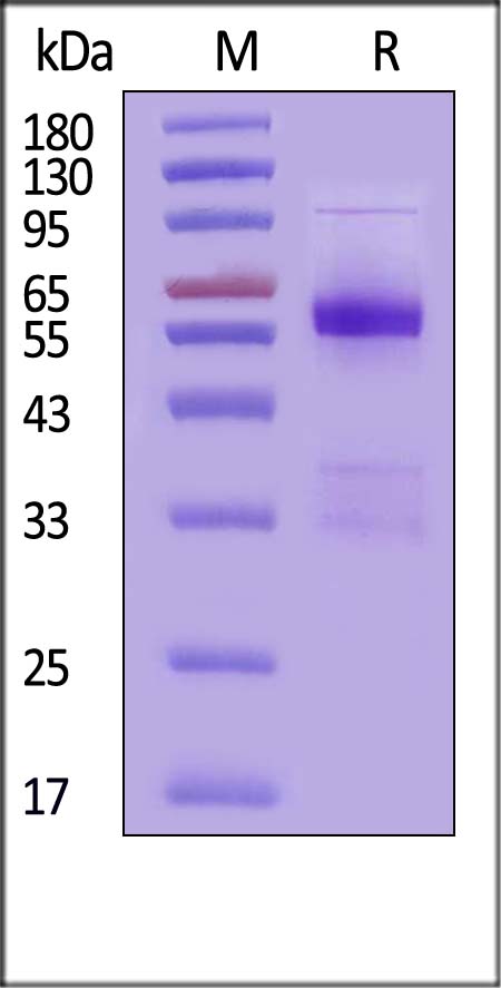
SDS-PAGE
Human USAG-1, Fc Tag on SDS-PAGE under reducing (R) condition. The gel was stained with Coomassie Blue. The purity of the protein is greater than 90% (With Star Ribbon Pre-stained Protein Marker).
Molecular Characteristics
Expression System: HEK293 (human 293 cells)
Protein Region: Phe24–Ser206
Predicted N-terminus: Phe24
Tag: Human IgG1 Fc (C-terminal)
Calculated Molecular Weight: 47.2 kDa
Due to glycosylation, the protein migrates at approximately:
33 kDa
38 kDa
55–65 kDa
~100 kDa (dimer)
when analyzed by SDS-PAGE under reducing conditions.
Purity & Quality Control
Purity: >90% (SDS-PAGE, Coomassie Blue staining)
Endotoxin Level: <1.0 EU/µg (LAL or rFC method)
Quality management compliant with ISO/GMP systems
Formulation
Lyophilized from a 0.22 μm filtered solution containing:
50 mM Tris
100 mM Glycine
25 mM Arginine
150 mM NaCl
pH 7.5
Trehalose (stabilizer)
Customized formulations available upon request.
Reconstitution & Storage
Reconstitution: Refer to the Certificate of Analysis (CoA) for detailed instructions.
Storage Conditions:
Lyophilized: −20°C to −70°C for up to 12 months
After reconstitution: −70°C for up to 3 months under sterile conditions
Avoid repeated freeze-thaw cycles
Biological Background
USAG-1 (SOSTDC1) is a secreted glycoprotein containing a C-terminal cysteine knot domain, a structural motif common in several growth factors including:
Transforming Growth Factor Beta (TGF-β)
Nerve Growth Factor (NGF)
Platelet-Derived Growth Factor (PDGF)
This cysteine knot structure plays a role in dimerization, receptor binding, and signal transduction.
USAG-1 acts as a negative regulator of BMP signaling, modulating:
Cellular proliferation
Differentiation
Apoptosis
Dentary morphogenesis
Endometrial implantation
Bone fracture healing
Additionally, SOSTDC1 participates in the regulation of the Wnt signaling pathway, implicating it in cancer biology and tissue development studies.
Applications
BMP signaling research
Wnt pathway studies
Bone biology research
Tooth and dentary development studies
Cancer signaling research
Receptor binding and Fc-fusion protein studies
Available Sizes
ETA for in-stock products: 4 business days
Safety Datasheet (SDS):
CLICK HERE TO VIEW SDS
Certificate of Analysis (COA):
To receive a Certificate of Analysis, please send us the lot number to info@dentinova.co.uk after purchasing.
Related Citations:
USAG-1 and Regenerative Dentistry, Therapeutic Implications and Future Directions: Review of the Literature Moradi, Karimi, Kolahdouz
et al Clin Exp Dent Res (2026) 12 (1), e70301
Abstract: Uterine Sensitization-Associated Gene 1 (USAG-1) is a bone morphogenetic protein (BMP) antagonist vital for tooth regeneration that is expressed in kidney, gingiva, and dental tissues.We analyzed recent studies focusing on USAG-1 and its involvement in BMP and Wnt signaling pathways related to dental tissue repair and regeneration. Preclinical models and clinical trial data were examined to evaluate the efficacy of USAG-1 inhibition as a therapeutic strategy. In addition, publicly available single-cell RNA sequencing and STRING databases were analyzed to investigate the gene expression of USAG-1 in human tissues and its protein interactions, respectively.RNA-seq analysis confirmed that USAG-1 is expressed in a subset of secretory cell types in kidney, jaw, and gingiva that are important for cell growth and morphogenesis. Recent studies have also demonstrated that inhibiting USAG-1 facilitates tooth regeneration by activating BMP-mediated morphogenesis and improving outcomes in preclinical models. Engineered monoclonal antibodies that target USAG-1 have shown that blocking the protein product of this gene can promote third dentition and alleviate congenital tooth agenesis. Clinical trials utilizing this antibody are currently underway, with prospects for commercial applications within the next decade. Despite these advancements, challenges related to safety, specificity, and delivery mechanisms remain.This review underscores the transformative potential of USAG-1-based therapies in regenerative dentistry, offering a paradigm shift in dental care by enabling biologically authentic tooth regeneration. However, the realization of these advancements in clinical practice requires overcoming significant barriers, including ensuring safety, optimizing delivery systems, and addressing ethical concerns. Continued interdisciplinary research is essential to fully harness the potential of USAG-1 in regenerative dentistry.© 2026 The Author(s). Clinical and Experimental Dental Research published by John Wiley & Sons Ltd.
Inhibition of USAG-1 improved delayed graft function in renal transplantation Luo, Ma, Sun
et al Transpl Immunol (2026) 94, 102348
Abstract: Delayed graft function (DGF) is a critical complication following kidney transplantation. This study aimed to identify novel biomarkers and therapeutic targets for DGF. Transcriptomic data from the Gene Expression Omnibus (GEO) database were analyzed to screen for DGF-associated core genes. Serum levels of uterine sensitization-associated gene-1 (USAG-1) and kidney injury molecule-1 (KIM-1) were measured one day post-transplantation, and their predictive values for DGF were compared. Mouse models of DGF were established by inducing graded warm ischemia. USAG-1 was identified as a core gene significantly associated with DGF. Multivariate logistic regression identified USAG-1 as an independent risk factor for DGF (P < 0.05). Importantly, USAG-1 demonstrated superior diagnostic performance with an area under the curve (AUC) of 0.774 (95% CI: 0.660-0.895), which outperformed the traditional marker KIM-1 (AUC = 0.686; 95% CI: 0.559-0.831) (Pcomparison < 0.05). High USAG-1 expression was significantly correlated with prolonged recovery time (P < 0.05) and post-transplant dialysis duration (P < 0.05). In mouse models, USAG-1 knockout (KO) significantly increased survival (P < 0.05), improved renal pathology, and reduced the levels of injury markers (all P < 0.01), whereas exogenous USAG-1 supplementation reversed these effects (all P < 0.05). In conclusion, USAG-1 is a key regulator in DGF pathogenesis. Early postoperative serum USAG-1 levels can effectively predict DGF, and inhibition of USAG-1 expression may alleviate renal injury and promote functional recovery, offering potential diagnostic and therapeutic value in renal transplantation.Copyright © 2026 The Authors. Published by Elsevier B.V. All rights reserved.
SOSTDC1 downregulation in CD4+ T cells confers protection against obesity-induced insulin resistance Li, Zhu, Zhang
et al Cell Rep (2025) 44 (4), 115496
Abstract: Adipose-resident T cells play a crucial role in the development of obesity-induced insulin resistance. However, the specific mechanisms, particularly those involving non-immune cytokines, remain unclear. Here, we report significantly elevated levels of sclerostin domain-containing protein 1 (SOSTDC1) in individuals with type 2 diabetes (T2D), showing positive correlations with fasting glucose and HbA1c. T cell-specific Sostdc1-deficient mice exhibit resistance to age-induced adipose lipid accumulation and glucose dysregulation at 12 months and protect against obesity-induced insulin resistance without affecting proinflammatory macrophage infiltration or adipose inflammation. Mechanistically, SOSTDC1 disrupts the lipid balance in adipocytes by promoting lipogenesis and inhibiting lipolysis through the LRP5/6-β-catenin pathway. Furthermore, T cell receptor (TCR) signaling significantly amplifies SOSTDC1 secretion in CD4+ T cells. In summary, our study uncovers an additional mechanism by which T cells contribute to obesity and insulin resistance, suggesting that inhibiting SOSTDC1 could be a promising immunotherapeutic strategy for metabolic disorders.Copyright © 2025 The Author(s). Published by Elsevier Inc. All rights reserved.
Development of a new antibody drug to treat congenital tooth agenesis Takahashi, Kiso, Mihara
et al J Oral Biosci (2024) 66 (4), 1-9
Abstract: This study aimed to develop a therapeutic agent promoting teeth regeneration from autologous tissues for congenital tooth agenesis, specifically for hypodontia (≤5 missing congenital teeth, 10% prevalence) and oligodontia (≥6 missing congenital teeth, 0.1% prevalence).We studied mice genetically deficient in the USAG-1 protein, an antagonist of BMP/Wnt which forms excessive teeth. We identified USAG-1 as a target molecule for increasing the number of teeth. Crossing USAG-1-deficient mice with a congenital tooth agenesis model restored tooth formation. We produced anti-USAG-1 neutralizing antibodies as potential therapeutic agents for the treatment of congenital tooth agenesis. Mice anti-USAG-1 neutralizing antibodies can potentially rescue the developmentally arrested tooth germ programmed to a certain tooth type. A humanized anti-USAG-1 antibody was developed as the final candidate.Targeting USAG-1 shows promise for treating missing congenital tooth. Anti-USAG-1 neutralizing antibodies have been developed and will progress towards clinical trials, which may regenerate missing congenital teeth in conditions, such as hypodontia and oligodontia. The protocol framework for a phase 1 study has been finalized, and preparation for future studies is underway.Copyright © 2024. Published by Elsevier B.V.
miR4352b a cross-species modulator of SOSTDC1, targets dual pathway to regulate bone health and fracture healing Rai, Sardar, Raj
et al Biochim Biophys Acta Mol Basis Dis (2025) 1871 (1), 167514
Abstract: Mutations in SOST can lead to various monogenic bone diseases. Its paralog, SOSTDC1, shares 55 % protein sequence homology and belongs to the BMP antagonist class. Sostdc1-/- mice exhibit distinct effects on cortical and trabecular bone. Genetic polymorphisms in SOSTDC1 impacting peak bone mass makes SOSTDC1 gene, a candidate for influencing BMD variation in humans. SOSTDC1 is upregulated in bone loss conditions, altering BMP-responsive genes and signaling modulators, suggesting its dual BMP/Wnt antagonist role may enhance both pathways. Overexpression of SOSTDC1 confirmed its role as an osteogenic antagonist. Glycine max (Soy)-derived miR4352b, identified for cross-kingdom applications, precisely targets SOSTDC1, a key regulator of bone. SOSTDC1 competitively binds to BMP2 receptor, BMPR1A. Gma-miR4352b suppresses SOSTDC1 expression, enhancing osteogenesis and countering SOSTDC1’s inhibition of osteogenic potential. Modeling estrogen deficiency to mimic elevated SOSTDC1 levels, we observed an inverse correlation with SOSTDC1 expression, while serum BMP2 and PINP levels increased following gma-miR4352b supplementation. In fracture healing, SOSTDC1’s crucial role becomes evident in conditions of delayed fracture healing. As healing progresses, SOSTDC1 expression decreases. Gma-miR4352b, compared to scrambled miRNA, remarkably promotes callus formation, achieving 68 % healing by day 10, surpassing the scrambled group at 44 %. By the day 13, the treatment group exhibits advanced healing, challenging to find the callus, while the scrambled group maintains a healing rate similar to day10. The accelerated healing in the treatment group underscores the importance of SOSTDC1 in influencing early fracture healing, potentially through the activation of both BMP2 and Wnt signaling pathways.Copyright © 2024 Elsevier B.V. All rights reserved.
SOSTDC1 Nuclear Translocation Facilitates BTIC Maintenance and CHD1-Mediated HR Repair to Promote Tumor Progression and Olaparib Resistance in TNBC Deng, Qiang, Liu
et al Adv Sci (Weinh) (2024) 11 (29), e2306860
Abstract: Breast tumor-initiating cells (BTICs) of triple-negative breast cancer (TNBC) tissues actively repair DNA and are resistant to treatments including chemotherapy, radiotherapy, and targeted therapy. Herein, it is found that a previously reported secreted protein, sclerostin domain containing 1 (SOSTDC1), is abundantly expressed in BTICs of TNBC cells and positively correlated with a poor patient prognosis. SOSTDC1 knockdown impairs homologous recombination (HR) repair, BTIC maintenance, and sensitized bulk cells and BTICs to Olaparib. Mechanistically, following Olaparib treatment, SOSTDC1 translocates to the nucleus in an importin-α dependent manner. Nuclear SOSTDC1 interacts with the N-terminus of the nucleoprotein, chromatin helicase DNA-binding factor (CHD1), to promote HR repair and BTIC maintenance. Furthermore, nuclear SOSTDC1 bound to β-transducin repeat-containing protein (β-TrCP) binding motifs of CHD1 is found, thereby blocking the β-TrCP-CHD1 interaction and inhibiting β-TrCP-mediated CHD1 ubiquitination and degradation. Collectively, these findings identify a novel nuclear SOSTDC1 pathway in regulating HR repair and BTIC maintenance, providing insight into the TNBC therapeutic strategies.© 2024 The Author(s). Advanced Science published by Wiley‐VCH GmbH.
Identification of genomic characteristics and selective signals in Guizhou black goat Chang, Zheng, Li
et al BMC Genomics (2024) 25 (1), 164
Abstract: Guizhou black goat is one of the indigenous black goat breeds in the southwest region of Guizhou, China, which is an ordinary goat for mutton production. They are characterized by moderate body size, black coat, favorite meat quality with tender meat and lower odor, and tolerance for cold and crude feed. However, little is known about the genetic characteristics or variations underlying their important economic traits.Here, we resequenced the whole genome of Guizhou black goat from 30 unrelated individuals breeding in the five core farms. A total of 9,835,610 SNPs were detected, and 2,178,818 SNPs were identified specifically in this breed. The population structure analysis revealed that Guizhou black goat shared a common ancestry with Shaanbei white cashmere goat (0.146), Yunshang black goat (0.103), Iran indigenous goat (0.054), and Moroccan goat (0.002). However, Guizhou black goat showed relatively higher genetic diversity and a lower level of linkage disequilibrium than the other seven goat breeds by the analysis of the nucleotide diversity, linkage disequilibrium decay, and runs of homozygosity. Based on FST and θπ values, we identified 645, 813, and 804 selected regions between Guizhou black goat and Yunshang black goat, Iran indigenous goat, and cashmere goats. Combined with the results of XP-EHH, there were 286, 322, and 359 candidate genes, respectively. Functional annotation analysis revealed that these genes are potentially responsible for the immune response (e.g., CD28, CD274, IL1A, TLR2, and SLC25A31), humility-cold resistance (e.g., HBEGF, SOSTDC1, ARNT, COL4A1/2, and EP300), meat quality traits (e.g., CHUK, GAB2, PLAAT3, and EP300), growth (e.g., GAB2, DPYD, and CSF1), fertility (e.g., METTL15 and MEI1), and visual function (e.g., PANK2 and NMNAT2) in Guizhou black goat.Our results indicated that Guizhou black goat had a high level of genomic diversity and a low level of linkage disequilibrium in the whole genome. Selection signatures were detected in the genomic regions that were mainly related to growth and development, meat quality, reproduction, disease resistance, and humidity-cold resistance in Guizhou black goat. These results would provide a basis for further resource protection and breeding improvement of this very local breed.© 2024. The Author(s).
Transcriptome analysis of adenomyosis eutopic endometrium reveals molecular mechanisms involved in adenomyosis-related implantation failure and pregnancy disorders Juárez-Barber, Corachán, Carbajo-García
et al Reprod Biol Endocrinol (2024) 22 (1), 10
Abstract: Women with adenomyosis are characterized by having defective decidualization, impaired endometrial receptivity and/or embryo-maternal communication, and implantation failure. However, the molecular mechanisms underlying adenomyosis-related infertility remain unknown, mainly because of the restricted accessibility and the difficult preservation of endometrial tissue in vitro. We have recently shown that adenomyosis patient-derived endometrial organoids, maintain disease-specific features while differentiated into mid-secretory and gestational endometrial phase, overcoming these research barriers and providing a robust platform to study adenomyosis pathogenesis and the associated molecular dysregulation related to implantation and pregnancy disorders. For this reason, we aim to characterize the dysregulated mechanisms in the mid-secretory and gestational endometrium of patients with adenomyosis by RNA-sequencing.Endometrial organoids were derived from endometrial biopsies collected in the proliferative phase of women with adenomyosis (ADENO) or healthy oocyte donors (CONTROL) (n = 15/group) and differentiated into mid-secretory (-SECorg) and gestational (-GESTorg) phases in vitro. Following RNA-sequencing, the significantly differentially expressed genes (DEGs) (FDR < 0.05) were identified and selected for subsequent functional enrichment analysis and QIAGEN Ingenuity Pathway Analysis (IPA). Statistical differences in gene expression were evaluated with the Student’s t-test or Wilcoxon test.We identified 1,430 DEGs in ADENO-SECorg and 1,999 DEGs in ADENO-GESTorg. In ADENO-SECorg, upregulated genes included OLFM1, FXYD5, and RUNX2, which are involved in impaired endometrial receptivity and implantation failure, while downregulated genes included RRM2, SOSTDC1, and CHAC2 implicated in recurrent implantation failure. In ADENO-GESTorg, upregulated CXCL14 and CYP24A1 and downregulated PGR were related to pregnancy loss. IPA predicted a significant inhibition of ID1 signaling, histamine degradation, and activation of HMGB1 and Senescence pathways, which are related to implantation failure. Alternatively, IPA predicted an inhibition of D-myo-inositol biosynthesis and VEGF signaling, and upregulation of Rho pathway, which are related to pregnancy loss and preeclampsia.Identifying dysregulated molecular mechanisms in mid-secretory and gestational endometrium of adenomyosis women contributes to the understanding of adenomyosis-related implantation failure and/or pregnancy disorders revealing potential therapeutic targets. Following experimental validation of our transcriptomic and in silico findings, our differentiated adenomyosis patient-derived organoids have the potential to provide a reliable platform for drug discovery, development, and personalized drug screening for affected patients.© 2024. The Author(s).
MiR-22-3p facilitates bone marrow mesenchymal stem cell osteogenesis and fracture healing through the SOSTDC1-PI3K/AKT pathway Wang, Wang, Cheng
et al Int J Exp Pathol (2024) 105 (2), 52-63
Abstract: Bone fractures are the most common form of musculoskeletal trauma worldwide. Numerous microRNAs (miRNAs) have been suggested to be participants in regulating bone-related diseases. Recent studies revealed the regulatory role of miR-22-3p in osteogenic differentiation, but its role in fracture healing has not been investigated previously. Here, a rat femoral fracture model was established, Bone marrow mesenchymal stem cells (BMSCs) were isolated to detect the specific function and underlying mechanisms of miR-22-3p. MiR-22-3p and sclerostin domain-containing 1 (SOSTDC1) expression was determined by RT-qPCR and immunohistochemistry staining. The levels of proteins associated with osteogenic differentiation were assessed by western blotting. Flow cytometry was conducted to identify the isolated rat BMSCs. Alizarin red staining, alkaline phosphatase staining and Oil Red O staining were used to evaluate the osteogenic and adipogenic differentiation of rat BMSCs. The interaction between miR-22-3p and SOSTDC1 was verified using a luciferase reporter assay. Haematoxylin and Eosin (H&E) staining of the bone tissues was performed to analyse the effect of miR-22-3p on histopathological changes in vivo. MiR-22-3p was downregulated in the callus tissues of rat femoral fracture, while the expression of SOSTDC1 was upregulated. The isolated rat BMSCs had the capacity for both osteogenic and adipogenic differentiation. The differentiation capacity of BMSCs into osteoblasts was increased by miR-22-3p overexpression. MiR-22-3p activated the PI3K/AKT pathway by targeting SOSTDC1. SOSTDC1 overexpression and PI3K/AKT signalling inhibitor LY294002 abolished the enhancing effect of miR-22-3p overexpression on the osteogenesis of BMSCs. Thus MiR-22-3p facilitated the femoral fracture healing in rats. MiR-22-3p overexpression promoted fracture healing via the activation of PI3K/AKT pathway by targeting SOSTDC1.© 2023 Company of the International Journal of Experimental Pathology (CIJEP).
Advances in tooth agenesis and tooth regeneration Ravi, Murashima-Suginami, Kiso
et al Regen Ther (2023) 22, 160-168
Abstract: The lack of treatment options for congenital (0.1%) and partial (10%) tooth anomalies highlights the need to develop innovative strategies. Over two decades of dedicated research have led to breakthroughs in the treatment of congenital and acquired tooth loss. We revealed that by inactivating USAG-1, congenital tooth agenesis can be successfully ameliorated during early tooth development and that the inactivation promotes late-stage tooth morphogenesis in double knockout mice. Furthermore, Anti- USAG-1 antibody treatment in mice is effective in tooth regeneration and can be a breakthrough in treating tooth anomalies in humans. With approximately 0.1% of the population suffering from congenital tooth agenesis and 10% of children worldwide suffering from partial tooth loss, early diagnosis will improve outcomes and the quality of life of patients. Understanding the role of pathogenic USAG-1 variants, their interacting gene partners, and their protein functions will help develop critical biomarkers. Advances in next-generation sequencing, mass spectrometry, and imaging technologies will assist in developing companion and predictive biomarkers to help identify patients who will benefit from tooth regeneration.© 2023 The Japanese Society for Regenerative Medicine. Production and hosting by Elsevier B.V.
Ageing-Associated Transcriptomic Alterations in Peri-Implantitis Pathology: A Bioinformatic Study Tian
Dis Markers (2022) 2022, 8456968
Abstract: Ageing is associated with increased incidence of peri-implantitis but the roles of ageing-associated biological mechanisms in the occurrence of peri-implantitis are not known. This study is aimed at performing integrative bioinformatic analysis of publically available datasets to uncover molecular mechanisms related to ageing and peri-implantitis.Gene expression datasets related to ageing and peri-implantitis (PI) were sought, and differentially expressed genes (DEGs) were analysed. Ageing-related genes were also identified from the “Aging Atlas” database. Using intersection analysis, an age-related-PI gene set was identified. Functional enrichment analysis for enriched GO biological process and KEGG pathways, protein-protein interaction (PPI) network analysis, correlation analysis, and immune cell infiltration analysis to determine high-abundance immune cells were performed. Least absolute shrinkage and selection operator (LASSO) logistic regression identified key age-related-PI genes. Transcription factor-gene and drug-gene interactions and enriched KEGG pathways for the key age-related-PI genes were determined.A total of 52 genes were identified as age-related-PI genes and found enriched in several inflammation-associated processes including myeloid leukocyte activation, acute inflammatory response, mononuclear cell differentiation, B cell activation, NF-kappa B signalling, IL-17 signalling, and TNF signalling. LYN, CDKN2A, MAPT, BTK, and PRKCB were hub genes in the PPI network. Immune cell infiltration analysis showed activated dendritic cells, central memory CD4 T cells, immature dendritic cells, and plasmacytoid dendritic cells were highly abundant in PI and ageing. 7 key age-related PI genes including ALOX5AP, EAF2, FAM46C, GZMK, MAPT, RGS1, and SOSTDC1 were identified using LASSO with high predictive values and found to be enriched in multiple neurodegeneration-associated pathways, MAPK signalling, and Fc epsilon RI signalling. MAPT and ALOX5AP were associated with multiple drugs and transcription factors and interacted with other age-related genes to regulate multiple biological pathways.A suite of bioinformatics analysis identified a 7-signature gene set highly relevant to cooccurrence of ageing and peri-implantitis and highlighted the role of neurodegeneration, autoimmune, and inflammation related pathways. MAPT and ALOX5AP were identified as key candidate target genes for clinical translation.Copyright © 2022 Zhaojun Tian.
SOSTDC1 acts as a tumor inhibitor in acute myeloid leukemia by downregulating the Wnt/β-catenin pathway Li, Wang, Shi
et al Environ Toxicol (2022) 37 (8), 1934-1943
Abstract: Sclerostin domain-containing 1 (SOSTDC1) has been documented as a key tumor-associated protein that is differentially expressed in multiple malignancies. However, the function of SOSTDC1 in acute myeloid leukemia (AML) is unexplored. The goal of this work was to assess the possible role of SOSTDC1 in AML. Our data showed decreased SOSTDC1 level in bone marrow from AML patients, and patients with low levels of SOSTDC1 had a reduced survival rate. SOSTC1 upregulation restrained the proliferative ability and promoted the apoptotic rate of AML cells. SOSTDC1 suppressed the activation of the Wnt/β-catenin pathway in AML cells. Reactivation of the Wnt/β-catenin pathway reversed SOSTDC1-mediated antitumor effects. SOSTDC1 upregulation weakened the tumorigenicity of AML cells in vivo. Collectively, our work demonstrates that SOSTDC1 has a tumor-inhibiting role in AML via downregulation of the Wnt/β-catenin pathway. This work underscores a key function for the SOSTDC1/Wnt/β-catenin pathway in AML.© 2022 Wiley Periodicals LLC.
USP15 negatively regulates lung cancer progression through the TRAF6-BECN1 signaling axis for autophagy induction Kim, Min, Jeong
et al Cell Death Dis (2022) 13 (4), 348
Abstract: TNF receptor-associated factor 6 (TRAF6)-BECN1 signaling axis plays a pivotal role in autophagy induction through ubiquitination of BECN1, thereby inducing lung cancer migration and invasion in response to toll-like receptor 4 (TLR4) stimulation. Herein, we provide novel molecular and cellular mechanisms involved in the negative effect of ubiquitin-specific peptidase 15 (USP15) on lung cancer progression. Clinical data of the TCGA and primary non-small cell lung cancer (NSCLC) patients (n = 41) revealed that the expression of USP15 was significantly downregulated in lung cancer patients. Importantly, USP15-knockout (USP15KO) A549 and USP15KO H1299 lung cancer cells generated with CRISPR-Cas9 gene-editing technology showed increases in cancer migration and invasion with enhanced autophagy induction in response to TLR4 stimulation. In addition, biochemical studies revealed that USP15 interacted with BECN1, but not with TRAF6, and induced deubiquitination of BECN1, thereby attenuating autophagy induction. Notably, in primary NSCLC patients (n = 4) with low expression of USP15, 10 genes (CCNE1, MMP9, SFN, UBE2C, CCR2, FAM83A, ETV4, MYO7A, MMP11, and GSDMB) known to promote lung cancer progression were significantly upregulated, whereas 10 tumor suppressor genes (FMO2, ZBTB16, FCN3, TCF21, SFTPA1B, HPGD, SOSTDC1, TMEM100, GDF10, and WIF1) were downregulated, providing clinical relevance of the functional role of USP15 in lung cancer progression. Taken together, our data demonstrate that USP15 can negatively regulate the TRAF6-BECN1 signaling axis for autophagy induction. Thus, USP15 is implicated in lung cancer progression.© 2022. The Author(s).
Exosomal circ_0001190 Regulates the Progression of Gastric Cancer via miR-586/SOSTDC1 Axis Liu, Yang, Zhu
et al Biochem Genet (2022) 60 (6), 1895-1913
Abstract: Gastric cancer (GC) is the fifth most common cancer, which has a significant impact on human health. Recent researches have shown that circular RNAs (circRNAs) could affect the progress of GC, but the mechanism still indistinct. In this work, we explored the roles of circ_0001190 in GC. The levels of circ_0001190, microRNA-586 (miR-586) and sclerostin domain containing 1 (SOSTDC1) were detected by quantitative RT-PCR and western blot in GC. The cell functions were scrutinized by cell counting kit-8 assay, 5-Ethynyl-29-deoxyuridine assay, flow cytometry assay, tube formation assay, transwell assay, and western blot. Furthermore, the relationship between miR-586 and circ_0001190 or SOSTDC1 was identified by dual-luciferase reporter assay. Finally, the xenograft model test was implemented to demonstrate the effect of exosomal circ_0001190 in vivo. The levels of circ_0001190 and SOSTDC1 were downregulated, and the miR-586 level was increased in GC. For functional assay, circ _0001190 overexpression inhibited cell vitality, cell proliferation, angiogenesis, cell migration and invasion, whereas stimulated cell apoptosis in GC cells. Circ _0001190 served as a miR-586 sponge to adjust the expression of SOSTDC1. Additionally, miR-586 could promote the advancement of GC by interfering SOSTDC1. Exosomal circ_0001190 overexpression inhibited the development of GC by miR-586/SOSTDC1 axis, which proposed a potential targeted therapy for GC cure.© 2022. The Author(s), under exclusive licence to Springer Science+Business Media, LLC, part of Springer Nature.
Proteomic profiling identifies novel diagnostic biomarkers and molecular subtypes for mucinous tubular and spindle cell carcinoma of the kidney Xu, Li, Zhu
et al J Pathol (2022) 257 (1), 53-67
Abstract: Mucinous tubular and spindle cell carcinoma (MTSCC) is a relatively rare renal epithelial neoplasm resembling type 1 papillary renal cell carcinoma (PRCC) morphologically and immunohistochemically. The accurate diagnosis of MTSCC remains a challenge. Here, by using proteomic profiling, we characterized MTSCC and PRCC to identify diagnostic biomarkers. We found that the MTSCC tumor proteome was significantly enriched in B-cell-mediated immunity when compared with the proteome of adjacent normal tissues of MTSCC or tumors of PRCC. Importantly, we identified MZB1, VCAN, and SOSTDC1 as diagnostic biomarkers to distinguish MTSCC from the solid variant of type 1 PRCC, with an AUC of 0.985 when combined. MZB1 was inversely correlated with tumor clinical stage and may play an anti-tumor role by activating the complement system. Finally, unsupervised clustering revealed two molecular subtypes of MTSCC, displaying different morphology, expression signatures of oxidative phosphorylation, and aggravation. In summary, our analyses identified a three-protein diagnostic panel and molecular subtypes for MTSCC. © 2022 The Authors. The Journal of Pathology published by John Wiley & Sons Ltd on behalf of The Pathological Society of Great Britain and Ireland.© 2022 The Authors. The Journal of Pathology published by John Wiley & Sons Ltd on behalf of The Pathological Society of Great Britain and Ireland.
Identification and validation of plasma biomarkers for diagnosis of breast cancer in South Asian women Rajkumar, Amritha, Sridevi
et al Sci Rep (2022) 12 (1), 100
Abstract: Breast cancer is the most common malignancy among women globally. Development of a reliable plasma biomarker panel might serve as a non-invasive and cost-effective means for population-based screening of the disease. Transcriptomic profiling of breast tumour, paired normal and apparently normal tissues, followed by validation of the shortlisted genes using TaqMan® Low density arrays and Quantitative real-time PCR was performed in South Asian women. Fifteen candidate protein markers and 3 candidate epigenetic markers were validated first in primary breast tumours and then in plasma samples of cases [N = 202 invasive, 16 DCIS] and controls [N = 203 healthy, 37 benign] using antibody array and methylation specific PCR. Diagnostic efficiency of single and combined markers was assessed. Combination of 6 protein markers (Adipsin, Leptin, Syndecan-1, Basic fibroblast growth factor, Interleukin 17B and Dickopff-3) resulted in 65% sensitivity and 80% specificity in detecting breast cancer. Multivariate diagnostic analysis of methylation status of SOSTDC1, DACT2, WIF1 showed 100% sensitivity and up to 91% specificity in discriminating BC from benign and controls. Hence, combination of SOSTDC1, DACT2 and WIF1 was effective in differentiating breast cancer [non-invasive and invasive] from benign diseases of the breast and healthy individuals and could help as a complementary diagnostic tool for breast cancer.© 2022. The Author(s).
Impact of microRNA-21-5p on the growth of thyroid cancer cells via targeting the recombinant sclerostin domain containing protein 1 Liu, Deng, Zhao
et al Zhong Nan Da Xue Xue Bao Yi Xue Ban (2021) 46 (10), 1054-1062
Abstract: To explore the molecular mechanism for thyroid cancer metastasis via analyzing the role of microRNA (miR)-21-5p and its target gene recombinant sclerostin domain containing protein 1 (SOSTDC1) in thyroid cancer.The target miR-21-5p was screened through bioinformatics analysis and cell verification, and the thyroid cancer cell lines was transfected with miR-21-5p inhibitor. 3-(4,5-dimethyl-2-thiazolyl)-2,5-diphenyl-2-H-tetrazolium bromide (MTT) test, flow cytometry, and cell scratch test were used to detect the proliferation, apoptosis and migration of thyroid cancer cells in the miR-21-5p inhibitor group and the inhibitor control group, respectively. The luciferase report experiment was used to verify the relationship between miR-21-5p and SOSTDC1, Western blotting was used to detect the expression levels and phosphorylation levels of SOSTDC1,phosphatidylinositol 3 kinase (PI3K), protein kinase B (Akt) and mitogen-activated protein kinases (MAPK), extracellular regulated protein kinases (ERK) in thyroid cancer cells.MiR-21-5p was significantly increased in thyroid cancer cells,which was negatively correlated with SOSTDC1 (r=-0.24, P<0.01). The proliferation and migration of thyroid cancer cells in the miR-21-5p inhibitor group was significantly lower than that in the inhibitor control group (both P<0.01), and the apoptosis rate in the miR-21-5p inhibitor group was significantly higher than that in the inhibitor control group (P<0.01).The luciferase report experiment showed that miR-21-5p could target and regulate the expression level of SOSTDC1, and the expression of PI3K in the miR-21-5p inhibitor group was significantly lower than that in the inhibitor control group (P<0.01). There were no significant changes in Akt and ERK1/2 levels, but the phosphorylation levels of Akt and ERK1/2 in the miR-21-5p inhibitor group were significantly lower than those in the inhibitor control group (both P<0.01).MiR-21-5p in thyroid cancer cells can target the expression of SOSTDC1 and affect the activities of PI3K/Akt and MAPK/ERK, thereby inhibiting the apoptosis of thyroid cancer cells and promoting cell proliferation and migration.
Evolution of the DAN gene family in vertebrates Opazo, Hoffmann, Zavala
et al Dev Biol (2022) 482, 34-43
Abstract: The DAN gene family (DAN, Differential screening-selected gene Aberrant in Neuroblastoma) is a group of genes that is expressed during development and plays fundamental roles in limb bud formation and digitation, kidney formation and morphogenesis and left-right axis specification. During adulthood the expression of these genes are associated with diseases, including cancer. Although most of the attention to this group of genes has been dedicated to understanding its role in physiology and development, its evolutionary history remains poorly understood. Thus, the goal of this study is to investigate the evolutionary history of the DAN gene family in vertebrates, with the objective of complementing the already abundant physiological information with an evolutionary context. Our results recovered the monophyly of all DAN gene family members and divide them into five main groups. In addition to the well-known DAN genes, our phylogenetic results revealed the presence of two new DAN gene lineages; one is only retained in cephalochordates, whereas the other one (GREM3) was only identified in cartilaginous fish, holostean fish, and coelacanth. According to the phyletic distribution of the genes, the ancestor of gnathostomes possessed a repertoire of eight DAN genes, and during the radiation of the group GREM1, GREM2, SOST, SOSTDC1, and NBL1 were retained in all major groups, whereas, GREM3, CER1, and DAND5 were differentially lost.Copyright © 2021 Elsevier Inc. All rights reserved.
Uterine Sensitization-Associated Gene-1 in the Progression of Kidney Diseases Li, Yue, Feng
et al J Immunol Res (2021) 2021, 9752139
Abstract: Uterine sensitization-associated gene-1 (USAG-1), originally identified as a secretory protein preferentially expressed in the sensitized rat endometrium, has been determined to modulate bone morphogenetic protein (BMP) and Wnt expression to play important roles in kidney disease. USAG-1 affects the progression of acute and chronic kidney damage and the recovery of allograft kidney function by regulating the BMP and Wnt signaling pathways. Moreover, USAG-1 has been found to be involved in the process of T cell immune response, and its ability to inhibit germinal center activity and reduce humoral immunity is of great significance for the treatment of autoimmune nephropathy and antibody-mediated rejection (AMR) after renal transplantation. This article summarizes the many advances made regarding the roles of USAG-1 in the progression of kidney disease and outlines potential treatments.Copyright © 2021 Xiaohu Li et al.
Local application of Usag-1 siRNA can promote tooth regeneration in Runx2-deficient mice Mishima, Takahashi, Kiso
et al Sci Rep (2021) 11 (1), 13674
Abstract: Runt-related transcription factor 2 (Runx2)-deficient mice can be used to model congenital tooth agenesis in humans. Conversely, uterine sensitization-associated gene-1 (Usag-1)-deficient mice exhibit supernumerary tooth formation. Arrested tooth formation can be restored by crossing both knockout-mouse strains; however, it remains unclear whether topical inhibition of Usag-1 expression can enable the recovery of tooth formation in Runx2-deficient mice. Here, we tested whether inhibiting the topical expression of Usag-1 can reverse arrested tooth formation after Runx2 abrogation. The results showed that local application of Usag-1 Stealth small interfering RNA (siRNA) promoted tooth development following Runx2 siRNA-induced agenesis. Additionally, renal capsule transplantation of siRNA-loaded cationized, gelatin-treated mouse mandibles confirmed that cationized gelatin can serve as an effective drug-delivery system. We then performed renal capsule transplantation of wild-type and Runx2-knockout (KO) mouse mandibles, treated with Usag-1 siRNA, revealing that hindered tooth formation was rescued by Usag-1 knockdown. Furthermore, topically applied Usag-1 siRNA partially rescued arrested tooth development in Runx2-KO mice, demonstrating its potential for regenerating teeth in Runx2-deficient mice. Our findings have implications for developing topical treatments for congenital tooth agenesis.
lncRNA CDKN2A-AS1 facilitates tumorigenesis and progression of epithelial ovarian cancer via modulating the SOSTDC1-mediated BMP-SMAD signaling pathway Zhao, Dong, Chu
et al Cell Cycle (2021) 20 (12), 1147-1162
Abstract: Ovarian cancer (OC) is the fifth most common female malignant tumor and the leading cause of cancer-related death in women worldwide. Epithelial ovarian cancer (EOC) is the predominant type of OC. Investigating the mechanism underlying tumorigenesis and progression of EOC is urgent. Our previous research has shown that long non-coding RNAs (lncRNAs) CDKN2A-AS1 is upregulated in EOC tissues and cells. Furthermore, we have predicted that CDKN2A-AS1 is associated with the bone morphogenetic protein (BMP)-SMAD signaling pathway, which is negatively regulated by the sclerostin domain containing 1 (SOSTDC1). Therefore, we conjecture that the CDKN2A-AS1 regulate BMP-SMAD signaling pathway via interacting with SOSTDC1, which need more investigation. Moreover, the functions of the BMP-SMAD signaling pathway and the SOSTDC1 on EOC are still unclear. Herein, we unearthed that CDKN2A-AS1, BMP2/4/7, SMAD1/5/9 and phosphorylation of SMAD1/5/9 (p-SMAD1/5/9) were upregulated in EOC tissues and cells, whereas SOSTDC1 was downregulated in EOC tissues and cells. We firstly demonstrated that CDKN2A-AS1 bound directly with the SOSTDC1. CDKN2A-AS1 downregulated the expression of SOSTDC1, but upregulated the expression of BMP2/4/7, SMAD1/5/9, and p-SMAD1/5/9. CDKN2A-AS1 promoted the proliferation, migration, invasion of EOC cells and tumor growth in vivo, whereas SOSTDC1 inhibited the proliferation, migration, invasion of EOC cells. Knockdown SOSTDC1 rescued the inhibitory effect of si-lncRNA CDKN2A-AS1 on the EOC cells proliferation, migration and invasion. These results demonstrated that CDKN2A-AS1activated the BMP-SMAD signaling pathway by directly bind with SOSTDC1 to promote EOC tumor growth. CDKN2A-AS1/SOSTDC1 axis may provide a novel therapeutic strategy for EOC treatment.
Cingulate networks associated with gray matter loss in Parkinson’s disease show high expression of cholinergic genes in the healthy brain Keo, Dzyubachyk, van der Grond
et al Eur J Neurosci (2021) 53 (11), 3727-3739
Abstract: Structural covariance networks are able to identify functionally organized brain regions by gray matter volume covariance across a population. We examined the transcriptomic signature of such anatomical networks in the healthy brain using postmortem microarray data from the Allen Human Brain Atlas. A previous study revealed that a posterior cingulate network and anterior cingulate network showed decreased gray matter in brains of Parkinson’s disease patients. Therefore, we examined these two anatomical networks to understand the underlying molecular processes that may be involved in Parkinson’s disease. Whole brain transcriptomics from the healthy brain revealed upregulation of genes associated with serotonin, GPCR, GABA, glutamate, and RAS-signaling pathways. Our results also suggest involvement of the cholinergic circuit, in which genes NPPA, SOSTDC1, and TYRP1 may play a functional role. Finally, both networks were enriched for genes associated with neuropsychiatric disorders that overlap with Parkinson’s disease symptoms. The identified genes and pathways contribute to healthy functions of the posterior and anterior cingulate networks and disruptions to these functions may in turn contribute to the pathological and clinical events observed in Parkinson’s disease.© 2021 The Authors. European Journal of Neuroscience published by Federation of European Neuroscience Societies and John Wiley & Sons Ltd.
Anti-USAG-1 therapy for tooth regeneration through enhanced BMP signaling Murashima-Suginami, Kiso, Tokita
et al Sci Adv (2021) 7 (7)
Abstract: Uterine sensitization-associated gene-1 (USAG-1) deficiency leads to enhanced bone morphogenetic protein (BMP) signaling, leading to supernumerary teeth formation. Furthermore, antibodies interfering with binding of USAG-1 to BMP, but not lipoprotein receptor-related protein 5/6 (LRP5/6), accelerate tooth development. Since USAG-1 inhibits Wnt and BMP signals, the essential factors for tooth development, via direct binding to BMP and Wnt coreceptor LRP5/6, we hypothesized that USAG-1 plays key regulatory roles in suppressing tooth development. However, the involvement of USAG-1 in various types of congenital tooth agenesis remains unknown. Here, we show that blocking USAG-1 function through USAG-1 knockout or anti-USAG-1 antibody administration relieves congenital tooth agenesis caused by various genetic abnormalities in mice. Our results demonstrate that USAG-1 controls the number of teeth by inhibiting development of potential tooth germs in wild-type or mutant mice missing teeth. Anti-USAG-1 antibody administration is, therefore, a promising approach for tooth regeneration therapy.Copyright © 2021 The Authors, some rights reserved; exclusive licensee American Association for the Advancement of Science. No claim to original U.S. Government Works. Distributed under a Creative Commons Attribution License 4.0 (CC BY).
Development of tooth regenerative medicine strategies by controlling the number of teeth using targeted molecular therapy Takahashi, Kiso, Murashima-Suginami
et al Inflamm Regen (2020) 40, 21
Abstract: Analysis of various genetically modified mice, with supernumerary teeth, has revealed the following two intrinsic molecular mechanisms that increase the number of teeth. One plausible explanation for supernumerary tooth formation is the rescue of tooth rudiments. Topical application of candidate molecules could lead to whole tooth formation under suitable conditions. Congenital tooth agenesis is caused by the cessation of tooth development due to the deletion of the causative gene and suppression of its function. The arrest of tooth development in Runx2 knockout mice, a mouse model of congenital tooth agenesis, is rescued in double knockout mice of Runx2 and Usag-1. The Usag-1 knockout mouse is a supernumerary model mouse. Targeted molecular therapy could be used to generate teeth in patients with congenital tooth agenesis by stimulating arrested tooth germs. The third dentition begins to develop when the second successional lamina is formed from the developing permanent tooth in humans and usually regresses apoptotically. Targeted molecular therapy, therefore, seems to be a suitable approach in whole-tooth regeneration by the stimulation of the third dentition. A second mechanism of supernumerary teeth formation involves the contribution of odontogenic epithelial stem cells in adults. Cebpb has been shown to be involved in maintaining the stemness of odontogenic epithelial stem cells and suppressing epithelial-mesenchymal transition. Odontogenic epithelial stem cells are differentiated from one of the tissue stem cells, enamel epithelial stem cells, and odontogenic mesenchymal cells are formed from odontogenic epithelial cells by epithelial-mesenchymal transition. Both odontogenic epithelial cells and odontogenic mesenchymal cells required to form teeth from enamel epithelial stem cells were directly induced to form excess teeth in adults. An approach for the development of targeted therapeutics has been the local application of monoclonal neutralizing antibody/siRNA with cationic gelatin for USAG-1 or small molecule for Cebpb.© The Author(s) 2020.
SOSTDC1 promotes invasion and liver metastasis in colorectal cancer via interaction with ALCAM/CD166 Bartolomé, Pintado-Berninches, Jaén
et al Oncogene (2020) 39 (38), 6085-6098
Abstract: The mechanistic basis of liver metastasis in colorectal cancer remains poorly understood. We previously reported that the sclerostin domain containing-1 (SOSTDC1) protein is overexpressed in the secretome of metastatic colorectal cancer cells and can inhibit liver homing. Here, we investigated the mechanisms of SOSTDC1 for promoting invasiveness and progression of colorectal cancer liver metastasis. SOSTDC1 inhibition of BMP4 maintains the expression of cancer stem cell traits, including SOX2 and NANOG. Immunoprecipitation and mass spectrometry analyses reveal the association of SOSTDC1 with ALCAM/CD166, which was confirmed by confocal microscopy and competition ELISA. Interaction with ALCAM is mediated by the N-terminal region of SOSTDC1, which contains a sequence similar to the ALCAM-binding motif used by CD6. Knocking down either SOSTDC1 or ALCAM expression, or using blocking antibodies, reduces the invasive activity by inhibiting Src and PI3K/AKT signaling pathways. In addition, ALCAM interacts with the α2ß1 and α1ß1 integrins, providing a possible link to Src activation. Finally, inoculation of SOSTDC1-silenced metastatic cells increases mouse survival by inhibiting liver metastasis. In conclusion, SOSTDC1 promotes invasion and liver metastasis in colorectal cancer, by overcoming BMP4-specific antimetastatic signals and inducing ALCAM-mediated Src and PI3K/AKT activation. These experiments underscore the potential of SOSTDC1 as a therapeutic target in metastatic colorectal cancer.
Characterization of the different oligomeric states of the DAN family antagonists SOSTDC1 and SOST Gipson, Kattamuri, Czepnik
et al Biochem J (2020) 477 (17), 3167-3182
Abstract: The DAN (differential screening-selected gene aberrative in neuroblastoma) family are a group of secreted extracellular proteins which typically bind to and antagonize BMP (bone morphogenetic protein) ligands. Previous studies have revealed discrepancies between the oligomerization state of certain DAN family members, with SOST (a poor antagonist of BMP signaling) forming a monomer while Grem1, Grem2, and NBL1 (more potent BMP antagonists) form non-disulfide linked dimers. The protein SOSTDC1 (Sclerostin domain containing protein 1) is sequentially similar to SOST, but has been shown to be a better BMP inhibitor. In order to determine the oligomerization state of SOSTDC1 and determine what effect dimerization might have on the mechanism of DAN family antagonism of BMP signaling, we isolated the SOSTDC1 protein and, using a battery of biophysical, biochemical, and structural techniques, showed that SOSTDC1 forms a highly stable non-covalent dimer. Additionally, this SOSTDC1 dimer was shown, using an in vitro cell based assay system, to be an inhibitor of multiple BMP signaling growth factors, including GDF5, while monomeric SOST was a very poor antagonist. These results demonstrate that SOSTDC1 is distinct from paralogue SOST in terms of both oligomerization and strength of BMP inhibition.© 2020 The Author(s).
Sostdc1 is expressed in all major compartments of developing and adult mammalian eyes Valensi, Goldman, Marchant
et al Graefes Arch Clin Exp Ophthalmol (2019) 257 (11), 2401-2427
Abstract: This study was conducted in order to study Sostdc1 expression in rat and human developing and adult eyes.Using the yeast signal sequence trap screening method, we identified the Sostdc1 cDNA encoding a protein secreted by the adult rat retinal pigment epithelium. We determined by in situ hybridization, RT-PCR, immunohistochemistry, and western blot analysis Sostdc1 gene and protein expression in developing and postnatal rat ocular tissue sections. We also investigated Sostdc1 immunohistolocalization in developing and adult human ocular tissues.We demonstrated a prominent Sostdc1 gene expression in the developing rat central nervous system (CNS) and eyes at early developmental stages from E10.5 days postconception (dpc) to E13 dpc. Specific Sostdc1 immunostaining was also detected in most adult cells of rat ocular tissue sections. We also identified the rat ocular embryonic compartments characterized by a specific Sostdc1 immunohistostaining and specific Pax6, Sox2, Otx2, and Vsx2 immunohistostaining from embryonic stages E10.5 to E13 dpc. Furthermore, we determined the localization of SOSTDC1 immunoreactivity in ocular tissue sections of developing and adult human eyes. Indeed, we detected SOSTDC1 immunostaining in developing and adult human retinal pigment epithelium (RPE) and neural retina (NR) as well as in several developing and adult human ocular compartments, including the walls of choroidal and scleral vessels. Of utmost importance, we observed a strong SOSTDC1 expression in a pathological ocular specimen of type 2 Peters’ anomaly complicated by retinal neovascularization as well in the walls ofother pathological extra-ocular vessels. CONCLUSION: As rat Sostdc1 and human SOSTDC1 are dual antagonists of the Wnt/β-catenin and BMP signaling pathways, these results underscore the potential crucial roles of these pathways and their antagonists, such as Sostdc1 and SOSTDC1, in developing and adult mammalian normal eyes as well as in syndromic and nonsyndromic congenital eye diseases.
Downregulation of Sostdc1 in Testicular Sertoli Cells is Prerequisite for Onset of Robust Spermatogenesis at Puberty Pradhan, Bhattacharya, Sarkar
et al Sci Rep (2019) 9 (1), 11458
Abstract: An alarming decline in sperm count of men from several countries has become a major concern for the world community. Hormones act on testicular Sertoli cells (Sc) to regulate male fertility by governing the division and differentiation of germ cells (Gc). However, there is a limited knowledge about Sc specific gene(s) regulating the spermatogenic output of the testis. Sclerostin domain-containing 1 protein (Sostdc1) is a dual BMP/Wnt regulator is predominantly expressed in the Sc of infant testes which hardly show any sign of spermatogenesis. In order to investigate the role of Sostdc1 in spermatogenic regulation, we have generated transgenic (Tg) rats which induced persistent expression of Sostdc1 in mature Sc causing reduced sperm counts. Although Sc specific Sostdc1 did not affect the function of either Sc or Leydig cells (Lc) in the adult testis of Tg rat, we observed a selective augmentation of the BMP target genes via activated phospho smad 1/5/8 signaling in Gc leading to apoptosis. Here, for the first time, we have demonstrated that Sostdc1 is a negative regulator of spermatogenesis, and provided substantial evidence that down regulation of Sostdc1 during puberty is critically essential for quantitatively and qualitatively normal spermatogenesis governing male fertility.
Lack of association between PAX6/SOSTDC1/FAM20B gene polymorphisms and mesiodens Liu, Li, Xu
et al BMC Oral Health (2019) 19 (1), 90
Abstract: The purpose of this study was to analyze the association between the genetic polymorphism of genes (PAX6, SOSTDC1and FAM20B) and the susceptibility to mesiodens.This study was carried out on 50 patients with mesiodens and 50 controls. The family history of each patient with mesiodens were recorded. Genomic DNA was extracted from saliva samples, and single nucleotide polymorphisms were detected in all exons and exon/intron boundaries of PAX6, SOSTDC1 and FAM20B using Sanger sequencing. The data were analyzed using pearson chi-square test with theoretical frequency ≥ 5. For theoretical frequency less than 5 but at least 1 (≤20% cell), the data were analyzed by continuity correction. For the rest, Fisher’s Exact test was used. A P-value< 0.05 was considered statistically significant. The Odds ratio (OR) and confidence intervals (CI) were recorded.Three polymorphisms were detected in PAX6. Two polymorphisms were detected in SOSTDC1. Twenty-nine polymorphisms were detected in FAM20B. Although, the T allele of FAM20B (rs3766626) appears to be associated with mesiodens (P = 0.051), there were no significant differences of PAX6/SOSTDC1/FAM20B gene polymorphisms between the two groups. The T allele of FAM20B (rs3766626) was associated with susceptibility to two mesiodens (P < 0.001; OR = 8.333; CI = 2.516-27.600).Lack of association between PAX6/SOSTDC1/FAM20B gene polymorphisms and mesiodens in the population studied was detected. Further studies with large samples on T allele of FAM20B (rs3766626) are needed.
Wnt Antagonists in Hematopoietic and Immune Cell Fate: Implications for Osteoporosis Therapies Chicana, Donham, Millan
et al Curr Osteoporos Rep (2019) 17 (2), 49-58
Abstract: We reviewed the current literature on the roles of the Wnt antagonists sclerostin (Sost) and sclerostin-containing domain protein 1 (Sostdc1) on bone homeostasis, the relationship of the hypoxia-inducible factor (Hif) and von Hippel-Lindau (Vhl) pathways on Sost expression, and how changes in bone induced by depletion of Sost, Sostdc1, and Vhl affect hematopoietic cells.B cell development is adversely affected in Sost-knockout mice and is more severely affected in Vhl-knockout mice. Inflammation in the Sost-/- bone microenvironment could alter hematopoietic stem cell behavior. Sostdc1-/- mice display defects in natural killer cell development and cytotoxicity. Depletion of Sost and Sostdc1 have effects on immune cell function that warrant investigation in patients receiving Wnt antagonist-depleting therapies for treatment of bone diseases. Additional clinical applications for manipulation of Wnt antagonists include cancer immunotherapies, stem cell transplantation, and directed differentiation to immune lineages.
Febuxostat inhibits TGF‑β1‑induced epithelial‑mesenchymal transition via downregulation of USAG‑1 expression in Madin‑Darby canine kidney cells in vitro Lu, Zhu, Zhang
et al Mol Med Rep (2019) 19 (3), 1694-1704
Abstract: Our previous study demonstrated that febuxostat, a xanthine oxidase inhibitor, can alleviate kidney dysfunction and ameliorate renal tubulointerstitial fibrosis in a rat unilateral ureteral obstruction (UUO) model; however, the underlying mechanisms remain unknown. Increasing evidence has revealed that epithelial‑mesenchymal transition (EMT) is one of the key mechanisms mediating the progression of renal tubulointerstitial fibrosis in chronic kidney disease (CKD). Uterine sensitization‑associated gene‑1 (USAG‑1), a kidney‑specific bone morphogenetic protein antagonist, is involved in the development of numerous types of CKDs. The present study aimed to investigate the role of febuxostat in the process of EMT in Madin‑Darby canine kidney (MDCK) cells in vitro. Western blotting, reverse transcription‑semiquantitative polymerase chain reaction analysis and immunofluorescence staining were used to evaluate the expression levels of bone morphogenetic protein 7, USAG‑1, α‑smooth muscle actin (α‑SMA) and E‑cadherin, respectively. The results demonstrated that the expression of USAG‑1 and α‑SMA increased, and that of E‑cadherin decreased significantly in MDCK cells following treatment with transforming growth factor‑β1 (TGF‑β1). The application of small interfering RNA‑USAG‑1 potently inhibited TGF‑β1‑induced EMT. Subsequently, the effects of febuxostat on TGF‑β1‑induced EMT was investigated. The results demonstrated that febuxostat downregulated the expression of USAG‑1, and reversed TGF‑β1‑induced EMT in MDCK cells. Furthermore, pretreatment with febuxostat significantly restored the decreased expression levels of phosphorylated Smad1/5/8 induced by TGF‑β1 in MDCK cells. The results of the present study suggested that USAG‑1 may be involved in the EMT process of MDCK cells induced by TGF‑β1, and febuxostat inhibited EMT by activating the Smad1/5/8 signaling pathway via downregulating the expression of USAG‑1 in MDCK cells.
Differential expression of secreted factors SOSTDC1 and ADAMTS8 cause profibrotic changes in linear morphoea fibroblasts Badshah, Brown, Weibel
et al Br J Dermatol (2019) 180 (5), 1135-1149
Abstract: Linear morphoea (LM) is a rare connective tissue disorder characterized by a line of thickened skin and subcutaneous tissue and can also affect the underlying muscle and bone. Little is known about the disease aetiology, with treatment currently limited to immune suppression, and disease recurrence post-treatment is common.In order to uncover new therapeutic avenues, the cell-intrinsic changes in LM fibroblasts compared with site-matched controls were characterized.We grew fibroblasts from site-matched affected and unaffected regions from five patients with LM, we subjected them to gene expression analysis and investigation of SMAD signalling.Fibroblasts from LM lesions showed increased migration, proliferation, altered collagen processing, and abnormally high basal levels of phosphorylated SMAD2, thereby rendering them less responsive to transforming growth factor (TGF)-β1 and reducing the degree of myofibroblast differentiation, which is a key component of the wound-healing and scarring process in normal skin. Conditioned media from normal fibroblasts could reverse LM-affected fibroblast migration and proliferation, suggesting that the LM phenotype is driven by an altered secretome. Gene array analysis and RNA-Seq indicated upregulation of ADAMTS8 and downregulation of FRAS1 and SOSTDC1. SOSTDC1 knock-down recapitulated the reduced TGF-β1 responsiveness and LM fibroblast migration, while overexpression of ADAMTS8 induced myofibroblast markers.We demonstrate that cell-intrinsic changes in the LM fibroblast secretome lead to changes observed in the disease, and that secretome modulation could be a viable therapeutic approach in the treatment of LM.© 2018 British Association of Dermatologists.
SOSTDC1 inhibits bone metastasis in non-small cell lung cancer and may serve as a clinical therapeutic target Chen, Gong, Wang
et al Int J Mol Med (2018) 42 (6), 3424-3436
Abstract: Bone metastasis occurs in ~40% patients with non‑small cell lung cancer (NSCLC), resulting in serious morbidity and mortality. Sclerostin domain‑containing protein 1 (SOSTDC1) has been demonstrated to be associated with the development and progression of multiple types of cancer. However, the role of SOSTDC1 in NSCLC bone metastasis remains unclear. In the present study, it was identified that SOSTDC1 was downregulated in NSCLC bone metastatic lesions compared with that in primary tumors, and low SOSTDC1 expression predicted poor prognosis for patients with NSCLC. Functionally, SOSTDC1 overexpression suppressed NSCLC cell proliferation, migration, invasion and cancer cell‑induced osteoclastogenesis, while SOSTDC1 knockdown produced the opposite effect. In addition, a number of potential downstream target genes of SOSTDC1, which were demonstrated to be associated with tumor progression and bone metastasis, were identified in NSCLC cells by RNA deep sequencing and RT‑qPCR assays. The results from the present study may provide useful insight for an improved understanding of the pathogenesis of NSCLC bone metastasis, and suggest that SOSTDC1 may be a potential prognostic biomarker and therapeutic target for NSCLC bone metastasis.
E4BP4 promotes thyroid cancer proliferation by modulating iron homeostasis through repression of hepcidin Zhou, Chen, Feng
et al Cell Death Dis (2018) 9 (10), 987
Abstract: Iron homeostasis is critical to mammals, and dysregulation in iron homeostasis usually leads to severe disorders including various cancers. Massive hepcidin secretion is an indicator of thyroid cancer, but the molecular mechanisms responsible for this dysregulation are unknown. Hepcidin secretion from thyroid cancer cells also leads to decreased expression of the iron exporter, ferroportin (FPN), and increased intracellular iron retention, which promote cancer proliferation. In this study, we examined the role of hepcidin in thyroid cancer (TC) and the molecular bases of its signaling. Synthesis of hepcidin is regulated by the BMP4/7 agonist SOSTDC1, which was downregulated in TC; SOSTDC1 downregulation was correlated with G9a-mediated hypermethylation in its promoter. The binding of G9a to the SOSTDC1 promoter requires E4BP4, which interacts with G9a to form a multi-molecular complex that contributes to SOSTDC1 silencing. Silencing of E4BP4 or G9a has similar effects to SOSTDC1 overexpression, which suppresses secretion of hepcidin and inhibits TC cell proliferation. Furthermore, our in vivo xenograft data indicated that depletion of E4BP4 also inhibits cancer growth, reduces hepcidin secretion, and reduces G9a nuclear transportation. Iron homeostasis and tumor growth in TC may be regulated by an E4BP4-dependent epigenetic mechanism. These findings suggest a new mechanism of cellular iron dysfunction through the E4BP4/G9a/SOSTDC1/hepcidin pathway, which is an essential link in TC.
Investigation of SOSTDC1 gene in non-syndromic patients with supernumerary teeth Arikan, Cumaogullari, Ozgul
et al Med Oral Patol Oral Cir Bucal (2018) 23 (5), e531-e539
Abstract: The etiology of supernumerary teeth is still unclear however heredity is believed to be a major factor and this idea was supported by several case reports. Recently, a relationship between supernumerary tooth formation and deficiency of Uterine Sensitization Associated Gene-1 (Usag-1), a rat gene that is expressed in sensitized endometrium, was reported in mice. The human homolog gene for Usag-1, Sclerostin Domain Containing 1 (SOSTDC1), shows 85% identity with mouse Usag-1. The present study aimed to investigate SOSTDC1 coding regions in non-syndromic patients with one or more supernumerary teeth.Twenty-five non-syndromic patients (21 male and 4 female) aged 5-15 years, with one or more supernumerary teeth were included in the study. Saliva samples were collected from patients and DNA samples were isolated and analyzed using PCR.Eight phenotypes of supernumerary tooth formation were observed in the study. From the DNA analysis, 2 novel and 3 previously identified sequence alterations were identified however, in investigating the Usag-1 homolog SOSTDC1 gene, the present study could not find any phenotype-genotype relationship.There are many SOSTDC1 homolog genes in the human genome and future studies should investigate these candidate genes. Also studies in larger case groups including family members may reveal the hereditary pattern.
Population-specific genetic modification of Huntington’s disease in Venezuela Chao, Kim, Shin
et al PLoS Genet (2018) 14 (5), e1007274
Abstract: Modifiers of Mendelian disorders can provide insights into disease mechanisms and guide therapeutic strategies. A recent genome-wide association (GWA) study discovered genetic modifiers of Huntington’s disease (HD) onset in Europeans. Here, we performed whole genome sequencing and GWA analysis of a Venezuelan HD cluster whose families were crucial for the original mapping of the HD gene defect. The Venezuelan HD subjects develop motor symptoms earlier than their European counterparts, implying the potential for population-specific modifiers. The main Venezuelan HD family inherits HTT haplotype hap.03, which differs subtly at the sequence level from European HD hap.03, suggesting a different ancestral origin but not explaining the earlier age at onset in these Venezuelans. GWA analysis of the Venezuelan HD cluster suggests both population-specific and population-shared genetic modifiers. Genome-wide significant signals at 7p21.2-21.1 and suggestive association signals at 4p14 and 17q21.2 are evident only in Venezuelan HD, but genome-wide significant association signals at the established European chromosome 15 modifier locus are improved when Venezuelan HD data are included in the meta-analysis. Venezuelan-specific association signals on chromosome 7 center on SOSTDC1, which encodes a bone morphogenetic protein antagonist. The corresponding SNPs are associated with reduced expression of SOSTDC1 in non-Venezuelan tissue samples, suggesting that interaction of reduced SOSTDC1 expression with a population-specific genetic or environmental factor may be responsible for modification of HD onset in Venezuela. Detection of population-specific modification in Venezuelan HD supports the value of distinct disease populations in revealing novel aspects of a disease and population-relevant therapeutic strategies.
Identification of methylation sites and signature genes with prognostic value for luminal breast cancer Xiao, Chen, Ke
et al BMC Cancer (2018) 18 (1), 405
Abstract: Robust and precise molecular prognostic predictors for luminal breast cancer are required. This study aimed to identify key methylation sites in luminal breast cancer, as well as precise molecular tools for predicting prognosis.We compared methylation levels of normal and luminal breast cancer samples from The Cancer Genome Atlas dataset. The relationships among differentially methylated sites, corresponding mRNA expression levels and prognosis were further analysed. Differentially expressed genes in normal and cancerous samples were analysed, followed by the identification of prognostic signature genes. Samples were divided into low- and high-risk groups based on the signature genes. Prognoses of low- and high-risk groups were compared. The Gene Expression Omnibus dataset were used to validate signature genes for prognosis prediction. Prognosis of low- and high-risk groups in Luminal A and Luminal B samples from the TCGA and the Metabric cohort dataset were analyzed. We also analysed the correlation between clinical features of low- and high- risk groups as well as their differences in gene expression.Fourteen methylation sites were considered to be related to luminal breast cancer prognosis because their methylation levels, mRNA expression and prognoses were closely related to each other. The methylation level of SOSTDC1 was used to divide samples into hypo- and hyper-methylation groups. We also identified an mRNA signature, comprising eight transcripts, ESCO2, PACSIN1, CDCA2, PIGR, PTN, RGMA, KLK4 and CENPA, which was used to divide samples into low- and high-risk groups. The low-risk group showed significantly better prognosis than the high-risk group. A correlation analysis revealed that the risk score was an independent prognostic factor. Low- and high- risk groups significantly correlated with the survival ratio in Luminal A samples, but not in Luminal B samples on the basis of the TCGA and the Metabric cohort dataset. Further functional annotation demonstrated that the differentially expressed genes were mainly involved in cell cycle and cancer progression.We identified several key methylation sites and an mRNA signature for predicting luminal breast cancer prognosis. The signature exhibited effective and precise prediction of prognosis and may serve as a prognostic and diagnostic marker for luminal breast cancer.
Regulation of CNKSR2 protein stability by the HECT E3 ubiquitin ligase Smurf2, and its role in breast cancer progression David, Surendran, Thulaseedharan
et al BMC Cancer (2018) 18 (1), 284
Abstract: Smurf2 E3 ubiquitin ligase physically associates with and regulate the stability of distinct cellular protein substrates. The multi-functional scaffold protein Connector enhancer of kinase suppressor of ras 2 (CNKSR2) plays a key role in regulating cell proliferation, and differentiation through multiple receptor tyrosine kinase pathways. The aim of this study was to investigate whether the interaction between Smurf2 and CNKSR2 has any significant role in the post transcriptional regulation of CNKSR2 expression in breast cancer.Here we demonstrate a novel interaction of CNKSR2 with Smurf2 by co-immunoprecipitation, indirect immunofluorescence studies, and surface plasmon resonance (SPR) analysis, which can ubiquitinate, but stabilize CNKSR2 by protecting it from proteasome mediated degradation.CNKSR2 protein levels were significantly increased upon forced overexpression of Smurf2, indicating the role of Smurf2 in regulating the stability of CNKSR2. Conversely, Smurf2 knockdown resulted in a marked decrease in the protein level expression of CNKSR2 by facilitating enhanced polyubiquitination and proteasomal degradation and reduced the proliferation and clonogenic survival of MDA-MB-231 breast cancer cell lines. Tissue microarray data from 84 patients with various stages of mammary carcinoma, including (in order of increasing malignant potential) normal, usual hyperplasia, fibrocystic changes, fibroadenoma, carcinoma-in-situ, and invasive ductal carcinoma showed a statistically significant association between Smurf2 and CNKSR2 expression, which is also well correlated with the ER, PR, and HER2 status of the tissue samples. A comparatively high expression of Smurf2 and CNKSR2 was observed when the expression of ER and PR was low, and HER2 was high. Consistently, both Smurf2 and CNKSR2 showed an integrated expression in MCF10 breast progression model cell lines.Altogether, our findings reveal that Smurf2 is a novel positive regulator of CNKSR2 and suggest that Smurf2-CNKSR2 interaction may serve as a common strategy to control proliferation of human breast cancer cells by modulating CNKSR2 protein stability.
Role of artesunate in TGF‑β1‑induced renal tubular epithelial‑mesenchymal transdifferentiation in NRK‑52E cells Zhang, Li, Zhu
et al Mol Med Rep (2017) 16 (6), 8891-8899
Abstract: The implications of epithelial‑mesenchymal transdifferentiation (EMT) have extended beyond the confines of renal fibrosis to renal tubulointerstitial fibrosis. It has been proposed that EMT may be one of the mechanisms involved in the pathogenesis of renal fibrosis. However, the underlying mechanisms remain unknown. Transforming growth factor (TGF)‑β1 is considered to be an important cytokine which regulates the transdifferentiation of tubular epithelial cells into myofibroblasts in renal tubulointerstitial fibrosis. In the present study, normal rat kidney tubular epithelial cells (NRK‑52E) were treated for 48 h with TGF‑β1 (5 ng/ml) and different concentrations of artesunate (ART; 0.01, 0.1 and 1 µg/ml). Western blotting, reverse transcription‑semi quantitative polymerase chain reaction analysis and immunofluorescence staining were used to evaluate the expression of bone morphogenetic protein (BMP)‑7, uterine sensitization‑associated gene (USAG)‑1, E‑cadherin, α‑smooth muscle actin (α‑SMA) and extracellular matrix collagen type I (Col I) mRNA. ART was able to attenuate renal injury in a unilateral ureteral obstruction model. However, its anti‑fibrotic effect remains to be elucidated. In the present study, it was observed that ART was able to ameliorate the TGF‑β1‑induced alterations in cellular morphology. In addition, ART inhibited the TGF‑β1‑induced USAG‑1 increase and the decrease in BMP‑7. Treatment with ART markedly attenuated the TGF‑β1‑induced upregulation of α‑SMA and downregulation of E‑cadherin. Additionally, ART was able to significantly attenuate the deposition of interstitial collagens, including Col I. The results of the present study further verified the therapeutic efficacy of ART in TGF‑β1‑induced renal interstitial fibrosis. These findings indicated that ART may hold the potential to prevent chronic kidney diseases via the suppression of USAG‑1 expression or by increasing BMP‑7 expression.
SOSTDC1 inhibits follicular thyroid cancer cell proliferation, migration, and EMT via suppressing PI3K/Akt and MAPK/Erk signaling pathways Zhou, Chen, Feng
et al Mol Cell Biochem (2017) 435 (1-2), 87-95
Abstract: The sclerostin domain containing protein 1 (SOSTDC1) is a cell signaling regulator involved in cell physiology and pathology. SOSTDC1 is known to have a suppressive effect on certain kinds of cancer. However, the role of SOSTDC1 in follicular thyroid cancer (FTC) remains unknown. We aimed to investigate if the expression of SOSTDC1 plays any roles in carcinogenesis and metastasis of FTC. We found a significantly down-regulated SOSTDC1 expression in follicular thyroid cancer samples. In addition, our data showed that ectopic expression of SOSTDC1 dramatically inhibited thyroid cancer cell proliferation in vitro and in nude mice. SOSTDC1 also compromised the migratory, invasive property, and epithelial-mesenchymal transition (EMT) activity of FTC cell. Mechanically, SOSTDC1 exerted its tumor suppressor function by inhibiting the activity of major signaling pathways including the phosphatidylinositol-3-kinase (PI3K)/Akt and mitogen-activated protein kinase (MAPK)/Erk pathways. Altogether, our findings provide insight into the role of SOSTDC1 as a novel functional tumor suppressor in follicular thyroid cancer through modulating the activities of PI3K/Akt and MAPK/Erk signaling pathways.
Biomarkers Predicting Bone Turnover in the Setting of CKD Evenepoel, Cavalier, D’Haese
Curr Osteoporos Rep (2017) 15 (3), 178-186
Abstract: Impaired bone quality contributes to the increased fracture risk in chronic kidney disease patients. Both low and high turnover bone disease may compromise bone quality. The question arises whether bone biomarkers may be additive or replace bone histormorphometry for diagnosing the extremes of bone turnover.Studies exploring the performance of established and emerging bone biomarkers against histomorphometric assessment of bone turnover are limited and overall yield inconclusive results as to their diagnostic utility. Bone biomarkers, although promising, currently fail to meet the needed diagnostic accuracy to replace bone histomorphometry and thus are not yet ready for clinical use. Bone biomarkers have not only several advantages, but also important limitations such as high biological variability, retention with kidney disease, preanalytical issues, and interassay variability. These important issues must be considered when developing and evaluating bone biomarkers. There is an urgent need for harmonization and standardization of available assays and additional bone biopsy studies.
Antagonistic Functions of USAG-1 and RUNX2 during Tooth Development Togo, Takahashi, Saito
et al PLoS One (2016) 11 (8), e0161067
Abstract: Supernumerary teeth and tooth agenesis are common morphological anomalies in humans. We previously obtained evidence that supernumerary maxillary incisors form as a result of the successive development of the rudimentary maxillary incisor tooth germ in Usag-1 null mice. The development of tooth germs is arrested in Runx2 null mice, and such mice also exhibit lingual epithelial buds associated with the upper molars and incisors. The aim of this study is to investigate the potential crosstalk between Usag-1 and Runx2 during tooth development. In the present study, three interesting phenomena were observed in double null Usag-1-/-/Runx2-/- mice: the prevalence of supernumerary teeth was lower than in Usag-1 null mice; tooth development progressed further compared than in Runx2 null mice; and the frequency of molar lingual buds was lower than in Runx2 null mice. Therefore, we suggest that RUNX2 and USAG-1 act in an antagonistic manner. The lingual bud was completely filled with odontogenic epithelial Sox2-positive cells in the Usag-1+/+/Runx2-/- mice, whereas almost no odontogenic epithelial Sox2-positive cells contributed to supernumerary tooth formation in the rudimentary maxillary incisors of the Usag-1-/-/Runx2+/+ mice. Our findings suggest that RUNX2 directly or indirectly prevents the differentiation and/or proliferation of odontogenic epithelial Sox2-positive cells. We hypothesize that RUNX2 inhibits the bone morphogenetic protein (BMP) and/or Wnt signaling pathways regulated by USAG-1, whereas RUNX2 expression is induced by BMP signaling independently of USAG-1.
Effects of Usag-1 and Bmp7 deficiencies on murine tooth morphogenesis Saito, Takahashi, Asahara
et al BMC Dev Biol (2016) 16, 14
Abstract: Wnt5a and Mrfzb1 genes are involved in the regulation of tooth size, and their expression levels are similar to that of Bmp7 during morphogenesis, including during the cap and early bell stages of tooth formation. We previously reported that Usag-1-deficient mice form supernumerary maxillary incisors. Thus, we hypothesized that BMP7 and USAG-1 signaling molecules may play important roles in tooth morphogenesis. In this study, we established double genetically modified mice to examine the in vivo inter-relationships between Bmp7 and Usag-1.We measured the volume and cross-sectional areas of the mandibular incisors using micro-computed tomography (micro-CT) in adult Bmp7- and Usag-1-LacZ knock-in mice and their F2 generation upon interbreeding. The mandibular incisors of adult Bmp7+/- mice were significantly larger than those of wild-type (WT) mice. The mandibular incisors of adult Usag-1-/- mice were the largest of all genotypes examined. In the F2 generation, the effects of these genes were additive; Bmp7+/- was most strongly associated with the increase in tooth size using generalized linear models, and the total area of mandibular supernumerary incisors of Usag-1-/-Bmp7+/- mice was significantly larger than that of Usag-1-/-Bmp7 +/+ mice. At embryonic day 15 (E15), BrdU assays demonstrated that the labeling index of Bmp7+/- embryos was significantly higher than that of WT embryos in the cervical loop. Additionally, the labeling index of Usag-1-/- embryos was significantly the highest of all genotypes examined in dental papilla.Bmp7 heterozygous mice exhibited significantly increased tooth sizes, suggesting that tooth size was controlled by specific gene expression. Our findings may be useful in applications of regenerative medicine and dentistry.
Sostdc1 deficiency accelerates fracture healing by promoting the expansion of periosteal mesenchymal stem cells Collette, Yee, Hum
et al Bone (2016) 88, 20-30
Abstract: Loss of Sostdc1, a growth factor paralogous to Sost, causes the formation of ectopic incisors, fused molars, abnormal hair follicles, and resistance to kidney disease. Sostdc1 is expressed in the periosteum, a source of osteoblasts, fibroblasts and mesenchymal progenitor cells, which are critically important for fracture repair. Here, we investigated the role of Sostdc1 in bone metabolism and fracture repair. Mice lacking Sostdc1 (Sostdc1(-/-)) had a low bone mass phenotype associated with loss of trabecular bone in both lumbar vertebrae and in the appendicular skeleton. In contrast, Sostdc1(-/-) cortical bone measurements revealed larger bones with higher BMD, suggesting that Sostdc1 exerts differential effects on cortical and trabecular bone. Mid-diaphyseal femoral fractures induced in Sostdc1(-/-) mice showed that the periosteal population normally positive for Sostdc1 rapidly expands during periosteal thickening and these cells migrate into the fracture callus at 3days post fracture. Quantitative analysis of mesenchymal stem cell (MSC) and osteoblast populations determined that MSCs express Sostdc1, and that Sostdc1(-/-) 5day calluses harbor >2-fold more MSCs than fractured wildtype controls. Histologically a fraction of Sostdc1-positive cells also expressed nestin and α-smooth muscle actin, suggesting that Sostdc1 marks a population of osteochondral progenitor cells that actively participate in callus formation and bone repair. Elevated numbers of MSCs in D5 calluses resulted in a larger, more vascularized cartilage callus at day 7, and a more rapid turnover of cartilage with significantly more remodeled bone and a thicker cortical shell at 21days post fracture. These data support accelerated or enhanced bone formation/remodeling of the callus in Sostdc1(-/-) mice, suggesting that Sostdc1 may promote and maintain mesenchymal stem cell quiescence in the periosteum.Copyright © 2016 The Authors. Published by Elsevier Inc. All rights reserved.
Artesunate attenuates unilateral ureteral obstruction-induced renal fibrosis by regulating the expressions of bone morphogenetic protein-7 and uterine sensitization-associated gene-1 in rats Cao, Wang, Li
et al Int Urol Nephrol (2016) 48 (4), 619-29
Abstract: Kidney fibrosis is the most common final stage of progressive renal disease. Bone morphogenetic protein-7 (BMP-7) has been shown to be important in both preservation of kidney function and resistance to injury. Recently, it has been realized that uterine sensitization-associated gene-1 (USAG-1) functions as a kidney-specific BMP antagonist. Because of the reported anti-fibrotic effects of artesunate (Art), this study was designed to investigate the effects of Art on renal fibrosis in unilateral ureteral obstruction (UUO) rats and to explore the underlying mechanisms.Thirty male Sprague-Dawley rats were randomly divided into five groups: Sham group, UUO group, low-dose Art-treated (Art-L), middle-dose Art-treated (Art-M), and high-dose Art-treated (Art-H) groups. The UUO rat model was established by ligating the left ureter. Fourteen days later, interstitial collagen deposition, expression of USAG-1, BMP-7, E-cadherin, α-smooth muscle actin (α-SMA), fibronectin (FN), collagen I, as well as the inflammatory infiltration levels in the kidneys were assessed.Art treatment significantly attenuated the deposition of interstitial collagens in the UUO rats’ kidneys and exhibited the ability to improve renal function, followed by the up-regulated expression of BMP-7 and E-cadherin and the down-regulated expression of USAG-1 and α-SMA. In addition, increased macrophages infiltration in the kidneys of the UUO rats were also attenuated by the administration of Art.These results indicate that Art is able to improve the renal function decline and renal fibrosis induced by UUO, which may be associated with the up-regulation of BMP-7 and down-regulation of USAG-1. Accordingly, Art may become a potential preventive or therapeutic agent for chronic kidney diseases.
Febuxostat Prevents Renal Interstitial Fibrosis by the Activation of BMP-7 Signaling and Inhibition of USAG-1 Expression in Rats Cao, Li, Peng
et al Am J Nephrol (2015) 42 (5), 369-78
Abstract: Renal interstitial fibrosis (RIF) is a common pathology associated with end-stage renal diseases. The activation of bone morphogenetic protein-7 (BMP-7)-Smad1/5/8 pathway seems to alleviate RIF. Uterine sensitization-associated gene-1 (USAG-1), a kidney-specific BMPs antagonist, is associated with the development and prognosis of several renal diseases. Febuxostat is a xanthine oxidase inhibitor that can attenuate the renal dysfunction of patients. The purpose of this study was to investigate the effects of febuxostat on renal fibrosis and to clarify the mechanisms underlying these effects.Rats were randomly divided into 6 groups termed a sham-operated group, a unilateral ureteral obstruction (UUO) group, 3 doses of febuxostat groups (low, intermediate and high doses) and a sham group treated with high-dose febuxostat. After 14 days, renal function, relative kidney weight, accumulation of glycogen and collagens were examined by different methods. Expression of α-SMA, transforming growth factor-β1 (TGF-β1), BMP-7 and USAG-1 was detected by western blotting and RT-PCR, respectively. The phosphorylation level of Smad1/5/8 was also quantified by western blotting.The renal function was declined, and large amounts of glycogen and collagens were deposited in the kidneys of UUO rats compared with the rats in the sham group. Besides, expression of α-SMA and USAG-1 in these kidneys was elevated, and the TGF-β1 was also activated, while the BMP-7-Smad1/5/8 pathway was inhibited. Febuxostat reversed the changes stated earlier, exhibiting protective effects on RIF induced by UUO.Febuxostat was able to attenuate RIF caused by UUO, which was associated with the activation of BMP-7-Smad1/5/8 pathway and the inhibition of USAG-1 expression in the kidneys of UUO rats.© 2015 S. Karger AG, Basel.
Effect of butyrate enemas on gene expression profiles and endoscopic/histopathological scores of diverted colorectal mucosa: A randomized trial Luceri, Femia, Fazi
et al Dig Liver Dis (2016) 48 (1), 27-33
Abstract: A temporary stoma is often created to protect a distal anastomosis in colorectal surgery. Short-chain fatty acids, mainly butyrate, are the major fuel source for the epithelium and their absence in the diverted tract may produce mucosal atrophy and inflammation.To investigate whether the administration of sodium butyrate enemas (Naburen(©), Promefarm, Italy) could prevent mucosal inflammation and atrophy and affect gene expression profiles after ileo/colostomy.We performed a randomized, double-blind, placebo-controlled clinical trial, in patients with enterostomy performed for inflammatory bowel disease, colorectal cancer or diverticulitis. Twenty patients were randomly allocated to receive 30ml of sodium butyrate 600mmol/L (group A) or saline (group B), b.i.d. for 30 days.In group A endoscopic scores were significantly improved (p<0.01) while mucosal atrophy was reduced or unchanged; in group B mucosal atrophy was increased in 42.8% of patients. Despite the high dose of butyrate used, no short-chain fatty acids were detectable by gas chromatography-mass spectrometry in colorectal biopsies. Group A patients showed up-regulation of genes associated with mucosal repair such as Wnt signalling, cytoskeleton regulation and bone morphogenetic protein-antagonists.Butyrate enemas may prevent the atrophy of the diverted colon/rectum, thus improving the recovery of tissue integrity.Copyright © 2015 Editrice Gastroenterologica Italiana S.r.l. Published by Elsevier Ltd. All rights reserved.
Down-regulation of SOSTDC1 promotes thyroid cancer cell proliferation via regulating cyclin A2 and cyclin E2 Liang, Guan, He
et al Oncotarget (2015) 6 (31), 31780-91
Abstract: Sclerostin domain containing protein 1 (SOSTDC1) is down-regulated and acts as a tumor suppressor in some kinds of cancers. However, the expression pattern and biological significance of SOSTDC1 in thyroid cancer are largely unknown. We demonstrated that SOSTDC1 was significantly down-regulated in thyroid cancer. Ectopic over-expression of SOSTDC1 inhibited proliferation and induced G1/S arrest in thyroid cancer cells. Moreover, SOSTDC1 over-expression suppressed the growth of tumor xenografts in nude mice. We also found that elevated SOSTDC1 led to inhibition of cyclin A2 and cyclin E2. Together,our results demonstrate that SOSTDC1 is down-regulated in thyroid cancer and might be a potential therapeutic target in the treatment of thyroid cancer.
A Small Molecule That Switches a Ubiquitin Ligase From a Processive to a Distributive Enzymatic Mechanism Kathman, Span, Smith
et al J Am Chem Soc (2015) 137 (39), 12442-5
Abstract: E3 ligases are genetically implicated in many human diseases, yet E3 enzyme mechanisms are not fully understood, and there is a strong need for pharmacological probes of E3s. We report the discovery that the HECT E3 Nedd4-1 is a processive enzyme and that disruption of its processivity by biochemical mutations or small molecules switches Nedd4-1 from a processive to a distributive mechanism of polyubiquitin chain synthesis. Furthermore, we discovered and structurally characterized the first covalent inhibitor of Nedd4-1, which switches Nedd4-1 from a processive to a distributive mechanism. To visualize the binding mode of the Nedd4-1 inhibitor, we used X-ray crystallography and solved the first structure of a Nedd4-1 family ligase bound to an inhibitor. Importantly, our study shows that processive Nedd4-1, but not the distributive Nedd4-1:inhibitor complex, is able to synthesize polyubiquitin chains on the substrate in the presence of the deubiquitinating enzyme USP8. Therefore, inhibition of E3 ligase processivity is a viable strategy to design E3 inhibitors. Our study provides fundamental insights into the HECT E3 mechanism and uncovers a novel class of HECT E3 inhibitors.
Hepcidin regulation in prostate and its disruption in prostate cancer Tesfay, Clausen, Kim
et al Cancer Res (2015) 75 (11), 2254-63
Abstract: Hepcidin is a circulating peptide hormone made by the liver that is a central regulator of systemic iron uptake and recycling. Here, we report that prostate epithelial cells also synthesize hepcidin, and that synthesis and secretion of hepcidin are markedly increased in prostate cancer cells and tissue. Prostatic hepcidin functions as an autocrine hormone, decreasing cell surface ferroportin, an iron exporter, increasing intracellular iron retention, and promoting prostate cancer cell survival. Synthesis of hepcidin in prostate cancer is controlled by a unique intersection of pathways that involves BMP4/7, IL6, Wnt, and the dual BMP and Wnt antagonist, SOSTDC1. Epigenetic silencing of SOSTDC1 through methylation is increased in prostate cancer and is associated with accelerated disease progression in patients with prostate cancer. These results establish a new connection between iron metabolism and prostate cancer, and suggest that prostatic dysregulation of hepcidin contributes to prostate cancer growth and progression.©2015 American Association for Cancer Research.
E4BP4 is a repressor of epigenetically regulated SOSTDC1 expression in breast cancer cells Rawat, Gopisetty, Thangarajan
Cell Oncol (Dordr) (2014) 37 (6), 409-19
Abstract: We and others show that SOSTDC1 is down-regulated in breast cancer tissues compared to matched normal tissues. Previously, we found that epigenetic mechanisms underlie the down-regulation of SOSTDC1 in gastric cancer cells. The aim of this study was to assess the putative epigenetic regulation of SOSTDC1 expression in breast cancer cells.Microarray-based expression profiling was performed in a series of primary breast cancers and matched normal tissues. Real-time PCR was performed to assess SOSTDC1 and E4BP4 mRNA levels in MCF7, BT549, MBMDA231, T47D (breast cancer) and HEK293T (normal kidney) cell lines. Methylation-specific PCR (MSP) and bisulfite sequencing PCR (BSP) were performed to assess the methylation level of the SOSTDC1 gene promoter, and 5-Aza 2-deoxycytidine (5′-Aza-dC) treatment was used to induce its demethylation. A luciferase assay was used to measure SOSTDC1 promoter activity in vitro. Stable shRNA-mediated knockdown of E4BP4 was carried out in MCF7 cells and confirmed by Western blotting. Finally, MCF7 cell proliferation and survival were measured by MTS assay.We found that SOSTDC1 is frequently down-regulated in primary breast cancers (98.2%) and in all breast cancer cell lines tested. MSP and BSP analyses revealed SOSTC1 promoter hypermethylation at CpG sites. 5′-Aza-dC treatment induced a striking down-regulation of SOSTDC1 gene expression, whereas BSP analysis showed demethylation of its promoter. Subsequent in silico SOSTDC1 promoter analysis indicated the presence of putative transcriptional repressor E4BP4 binding sites, and promoter deletion studies indeed revealed repressor binding regions encompassing these E4BP4 binding sites. Relative quantification of E4BP4 expression showed an inverse correlation to SOSTDC1 expression in the breast cancer cell lines tested. Exogenous over-expression of E4BP4 in HEK-293 and BT549 cells reduced SOSTDC1 expression and its promoter activity, respectively. Stable shRNA-mediated E4BP4 BT549 and MCF7 knock-down cells treated with 5′-Aza-dC exhibited up-regulation of SOSTDC1 expression and a concomitant inhibition of cell proliferation and survival.From our results we conclude that the transcriptional repressor E4BP4 plays a role in repressing epigenetically regulated SOSTDC1 expression in breast cancer cells, which can be reverted by E4BP4 silencing.
Interactions between BMP-7 and USAG-1 (uterine sensitization-associated gene-1) regulate supernumerary organ formations Kiso, Takahashi, Saito
et al PLoS One (2014) 9 (5), e96938
Abstract: Bone morphogenetic proteins (BMPs) are highly conserved signaling molecules that are part of the transforming growth factor (TGF)-beta superfamily, and function in the patterning and morphogenesis of many organs including development of the dentition. The functions of the BMPs are controlled by certain classes of molecules that are recognized as BMP antagonists that inhibit BMP binding to their cognate receptors. In this study we tested the hypothesis that USAG-1 (uterine sensitization-associated gene-1) suppresses deciduous incisors by inhibition of BMP-7 function. We learned that USAG-1 and BMP-7 were expressed within odontogenic epithelium as well as mesenchyme during the late bud and early cap stages of tooth development. USAG-1 is a BMP antagonist, and also modulates Wnt signaling. USAG-1 abrogation rescued apoptotic elimination of odontogenic mesenchymal cells. BMP signaling in the rudimentary maxillary incisor, assessed by expressions of Msx1 and Dlx2 and the phosphorylation of Smad protein, was significantly enhanced. Using explant culture and subsequent subrenal capsule transplantation of E15 USAG-1 mutant maxillary incisor tooth primordia supplemented with BMP-7 demonstrated in USAG-1+/- as well as USAG-1-/- rescue and supernumerary tooth development. Based upon these results, we conclude that USAG-1 functions as an antagonist of BMP-7 in this model system. These results further suggest that the phenotypes of USAG-1 and BMP-7 mutant mice reported provide opportunities for regenerative medicine and dentistry.
Vitamin D receptor-mediated control of Soggy, Wise, and Hairless gene expression in keratinocytes Hsieh, Estess, Kaneko
et al J Endocrinol (2014) 220 (2), 165-78
Abstract: The vitamin D receptor (VDR), but not its hormonal ligand, 1,25-dihydroxyvitamin D3 (1,25D), is required for the progression of the mammalian hair cycle. We studied three genes relevant to hair cycle signaling, DKKL1 (Soggy), SOSTDC1 (Wise), and HR (Hairless), to determine whether their expression is regulated by VDR and/or its 1,25D ligand. DKKL1 mRNA was repressed 49-72% by 1,25D in primary human and CCD-1106 KERTr keratinocytes; a functional vitamin D responsive element (VDRE) was identified at -9590 bp in murine Soggy. Similarly, SOSTDC1 mRNA was repressed 41-59% by 1,25D in KERTr and primary human keratinocytes; a functional VDRE was located at -6215 bp in human Wise. In contrast, HR mRNA was upregulated 1.56- to 2.77-fold by 1,25D in primary human and KERTr keratinocytes; a VDRE (TGGTGAgtgAGGACA) consisting of an imperfect direct repeat separated by three nucleotides (DR3) was identified at -7269 bp in the human Hairless gene that mediated dramatic induction, even in the absence of 1,25D ligand. In parallel, a DR4 thyroid hormone responsive element, TGGTGAggccAGGACA, was identified at +1304 bp in the human HR gene that conferred tri-iodothyronine (T3)-independent transcriptional activation. Because the thyroid hormone receptor controls HR expression in the CNS, whereas VDR functions in concert with the HR corepressor specifically in skin, a model is proposed wherein unliganded VDR upregulates the expression of HR, the gene product of which acts as a downstream comodulator to feedback-repress DKKL1 and SOSTDC1, resulting in integration of bone morphogenic protein and Wnt signaling to drive the mammalian hair cycle and/or influencing epidermal function.
Effects of estrogen on bone mRNA levels of sclerostin and other genes relevant to bone metabolism in postmenopausal women Fujita, Roforth, Demaray
et al J Clin Endocrinol Metab (2014) 99 (1), E81-8
Abstract: Studies in postmenopausal women have shown that estrogen reduces circulating sclerostin levels, but effects of estrogen on skeletal sclerostin mRNA levels are unknown.The objective of the study was to evaluate the effects of short-term estrogen treatment on bone mRNA levels of sclerostin and other genes relevant to bone metabolism.Needle bone biopsies were obtained from 20 postmenopausal women treated with transdermal estrogen for 3 weeks and 20 untreated controls. Quantitative PCR analyses were used to examine the expression of sclerostin and other genes related to bone metabolism, including 71 additional genes linked to bone density/fracture from genome-wide association studies.Estrogen treatment was associated with lower bone sclerostin mRNA levels (by 48%, P<.05) and with lower (by 54%, P<.01) mRNA levels of the sclerostin-related protein, sclerostin domain-containing protein 1 (SOSTDC1), which is also a Wnt/bone morphogenetic protein inhibitor. Consistent with studies in mice showing that ovariectomy increased nuclear factor-κB (NF-κB) activation, we found that estrogen treatment was associated with a significant reduction in inflammatory genes as a group (P=.028), with bone mRNA levels of NFKB2 and RELB (both encoding proteins in the NF-κB transcription factor complex) being significantly reduced individual genes. Eight of the 71 genome-wide association study-related genes examined were modulated by estrogen (P<.05, false discovery rate<0.10).In humans, estrogen-induced decreases in two key inhibitors of Wnt/bone morphogenetic protein signaling, sclerostin and SOSTDC1, along with reductions in NF-κB signaling, may be responsible for at least part of the protective effects of estrogen on bone.
SOSTDC1 down-regulation of expression involves CpG methylation and is a potential prognostic marker in gastric cancer Gopal, Raja, Shirley
et al Cancer Genet (2013) 206 (5), 174-82
Abstract: Sclerostin domain containing 1 (SOSTDC1) is reportedly down-regulated in various cancers. Our purpose was to study whether epigenetic mechanisms were involved in the down-regulation of expression in gastric cancer. Expression analysis of SOSTDC1 in gastric cancer cell lines indicated mRNA down-regulation. Our reporter assays and gene reactivation studies using 5-aza-2′-deoxycytidine, a DNA demethylating agent, and trichostatin A (TSA), a histone deacetylase (HDAC) inhibitor, demonstrated that epigenetic mechanisms are involved in the down-regulation of SOSTC1 expression. Methylation analysis of the SOSTDC1 promoter CpGs using methylation-specific polymerase chain reaction analysis revealed methylation in gastric cancer cell lines and tissue samples. A majority of tumors (17 of 18) with observed methylation exhibited down-regulation of mRNA expression relative to apparently normal gastric tissues. Immunoreactivity for SOSTDC1 in gastric tumors (24 of 46, 52.1%) was down-regulated relative to normal tissues (36 of 38, 94.7%) (P = 0.00001). The difference in expression between gastric tumor subtypes, intestinal and diffuse, was significant (P = 0.040). Expression of SOSTDC1 in gastric tumors increased the probability of both overall and disease-free survival. When overexpressed in AGS cells, cell proliferation, cell cycle progression, and anchorage-independent growth was repressed. The present findings indicate SOSTDC1 down-regulation involves methylation; SOSTDC1 expression is a potential prognostic factor and tumor suppressor in gastric cancer.Copyright © 2013 Elsevier Inc. All rights reserved.
3-Hydroxy-3-methylglutaryl-coenzyme A reductase inhibitor simvastatin ameliorates renal fibrosis through HOXA13-USAG-1 pathway Hamasaki, Doi, Okamoto
et al Lab Invest (2012) 92 (8), 1161-70
Abstract: Epidemiological data have suggested that 3-hydroxy-3-methylglutaryl-coenzyme A reductase inhibitors (statins) prevent the progression of chronic kidney diseases (CKDs), whereas the precise mechanism explaining in vitro to in vivo is missing. This study is aimed at exploring a new mechanism of action by statins on renal fibrosis, a hallmark of CKD, using mouse renal fibrosis model in vivo and Madin-Darby canine kidney (MDCK) cells expressing USAG-1 in vitro. C57/BL6 mice fed a 0.2% adenine-containing diet for 4 weeks developed renal dysfunction accompanied with severe tubulointerstitial fibrosis. Subsequent simvastatin (SIM) treatment (50 mg/kg per day) for 2 weeks significantly suppressed fibrosis progression. We found that SIM enhanced bone morphogenetic protein-7 (BMP-7)-mediated anti-fibrotic signaling with the reduced expression of uterine sensitization-associated gene-1 (USAG-1), a BMP-7 antagonist produced by renal distal tubular epithelial cells. Therefore, MDCK cells were incubated with transforming growth factor-β1 and showed increased expression of USAG-1 and α-smooth muscle actin; SIM significantly reduced them. SIM significantly increased E-cadherin expression. Gene knockdown experiments using MDCK suggested that homeobox protein Hox-A13 (HOXA13) played a suppressive role in the USAG-1 gene and thus SIM reduced USAG-1 by increasing HOXA13 expression. The data from our study demonstrate that SIM, one of statins, contributes to prevent the progression of renal fibrosis by upregulating BMP-7-mediated anti-fibrotic signaling and that one aspect of crucial efficacies is achieved by regulating HOXA13 and USAG-1. HOXA13-USAG-1 pathway is a newly identified mechanism in renal fibrosis and will be a new therapeutic target for preventing renal fibrosis progression in CKDs.
Selecting highly structure-specific antibodies using structured synthetic mimics of the cystine knot protein sclerostin Back, Frisch, Van Pee
et al Protein Eng Des Sel (2012) 25 (5), 251-9
Abstract: Antibodies directed against specific regions of a protein have traditionally been raised against full proteins, protein domains or simple unstructured peptides, containing contiguous stretches of primary sequence. We have used a new approach of selecting antibodies against restrained peptides mimicking defined epitopes of the bone modulator protein sclerostin, which has been identified as a negative regulator of the Wnt pathway. For a fast exploration of activity defining epitopes, we produced a set of synthetic peptide constructs mimicking native sclerostin, in which intervening loops from the cystine-knot protein sclerostin were truncated and whose sequences were optimized for fast and productive refolding. We found that the second loop within the cystine knot could be replaced by unnatural sequences, both speeding up folding, and increasing yield. Subsequently, we used these constructs to pan the HuCAL phage display library for antibodies capable of binding the native protein, thereby restricting recognition to the desired epitope regions. It is shown that the antibodies that were obtained recognize a complex epitope in the protein that cannot be mimicked with linear peptides. Antibodies selected against peptides show similar recognition specificity and potency as compared with antibodies obtained from full-length recombinant protein.
Bmp modulators in kidney disease Nakamura, Yanagita
Discov Med (2012) 13 (68), 57-63
Abstract: Bone morphogenetic proteins (Bmps) are phylogenetically conserved signaling molecules that belong to the transforming growth factor beta superfamily. Although these proteins were first identified because of their ability to induce ectopic bone and cartilage formation, they are also involved in the cascades of body patterning and morphogenesis. Recently, several reports have indicated that the administration of pharmacological doses of Bmps inhibits and repairs acute and chronic renal injury in animal models. However, its mechanism of action and physiological function are not well understood. In addition, the exogenous administration of Bmps causes undesired side effects in other tissues, because Bmp receptors are widely expressed. The activities of Bmps are regulated by Bmp antagonists, which bind directly to Bmp and inhibit its binding to the receptor. Thus, the Bmp antagonists that modulate endogenous Bmp activities may be possible new therapeutic targets for kidney disease. In this review, we discuss recent findings related to Bmp antagonists modifying the function of Bmps in kidney disease.
Contribution of the sclerostin domain-containing protein 1 (SOSTDC1) gene to normal variation of peak bone mineral density in Chinese women and men He, Yue, Hu
et al J Bone Miner Metab (2011) 29 (5), 571-81
Abstract: A genome-wide linkage analysis in Chinese families revealed a significant quantitative trait loci on chromosome 7p21.1 for femoral neck bone mineral density (BMD) (LOD = 3.68), and a potential candidate gene, sclerostin domain-containing protein 1 (SOSTDC1), is located in this region. SOSTDC1 belongs to a class of bone morphogenetic protein (BMP) antagonists that bind BMPs and regulate their signaling. We therefore genotyped 6 tag single nucleotide polymorphisms (tag-SNPs) in SOSTDC1 gene using allele-specific PCR method and investigated the association between SOSTDC1 gene polymorphisms and peak BMD variation in 401 Chinese female-offspring nuclear families (including 1260 subjects) and 400 Chinese male-offspring nuclear families (including 1215 subjects), respectively. Using both family-based (quantitative transmission disequilibrium test) and population-based (ANOVA) methods of analyses, BMD values were adjusted for age, height and weight. In female-offspring nuclear families, we found a significant within family association between rs16878759 and the lumbar spine peak BMD (P = 0.003) and rs16878759 accounted for 1.4% of the lumbar spine peak BMD variation. Moreover, haplotype CCC (containing rs12699800, rs16878759, and rs17619769) had a significant within family association with the lumbar spine peak BMD (P = 0.001) and accounted for 1.9% of the peak BMD variation at this bone site. However, in the male-offspring nuclear families, we failed to detect any significant association between any SNP or haplotype and peak BMD at any bone site. In conclusion, our results indicate for the first time that the genetic polymorphisms in SOSTDC1 have an effect on attainment and maintenance of peak bone mass in Chinese women.
SOSTDC1 differentially modulates Smad and beta-catenin activation and is down-regulated in breast cancer Clausen, Blish, Birse
et al Breast Cancer Res Treat (2011) 129 (3), 737-46
Abstract: Sclerostin domain containing 1 (SOSTDC1) protein regulates processes from development to cancer by modulating activity of bone morphogenetic protein (BMP) and wingless/int (Wnt) signaling pathways. As dysregulation of both BMP and Wnt signaling has been observed in breast cancer, we investigated whether disruption of SOSTDC1 signaling occurs in breast cancer. SOSTDC1 mRNA expression levels in breast tissue were examined using a dot blot. Affymetrix microarray data on SOSTDC1 levels were correlated with breast cancer patient survival using Kaplan-Meier plots. Correlations between SOSTDC1 protein levels and clinical parameters were assessed by immunohistochemistry of a breast cancer tissue microarray. SOSTDC1 secretion and BMP and Wnt signaling were investigated using immunoblotting. We found that SOSTDC1 is expressed in normal breast tissue and this expression is reduced in breast cancer. High levels of SOSTDC1 mRNA correlated with increased patient survival; conversely, SOSTDC1 protein levels decreased as tumor size and disease stage increased. Treatment of breast cancer cells with recombinant SOSTDC1 or Wise, a SOSTDC1 orthologue, demonstrated that SOSTDC1 selectively blocks BMP-7-induced Smad phosphorylation without diminishing BMP-2 or Wnt3a-induced signaling. In conclusion, SOSTDC1 mRNA and protein are reduced in breast cancer. High SOSTDC1 mRNA levels correlate with increased distant metastasis-free survival in breast cancer patients. SOSTDC1 differentially affects Wnt3a, BMP-2, and BMP-7 signaling in breast cancer cells. These results identify SOSTDC1 as a clinically important extracellular regulator of multiple signaling pathways in breast cancer.
Loss of heterozygosity and SOSTDC1 in adult and pediatric renal tumors Blish, Clausen, Hawkins
et al J Exp Clin Cancer Res (2010) 29 (1), 147
Abstract: Deletions within the short arm of chromosome 7 are observed in approximately 25% of adult and 10% of Wilms pediatric renal tumors. Within Wilms tumors, the region of interest has been delineated to a 2-Mb minimal region that includes ten known genes. Two of these ten candidate genes, SOSTDC1 and MEOX2, are particularly relevant to tumor development and maintenance. This finding, coupled with evidence that SOSTDC1 is frequently downregulated in adult renal cancer and regulates both Wingless-Int (Wnt)- and bone morphogenetic protein (BMP)-induced signaling, points to a role for SOSTDC1 as a potential tumor suppressor.To investigate this hypothesis, we interrogated the Oncomine database to examine the SOSTDC1 levels in adult renal clear cell tumors and pediatric Wilms tumors. We then performed single nucleotide polymorphism (SNP) and sequencing analyses of SOSTDC1 in 25 pediatric and 36 adult renal tumors. Immunohistochemical staining of patient samples was utilized to examine the impact of SOSTDC1 genetic aberrations on SOSTDC1 protein levels and signaling.Within the Oncomine database, we found that SOSTDC1 levels were reduced in adult renal clear cell tumors and pediatric Wilms tumors. Through SNP and sequencing analyses of 25 Wilms tumors, we identified four with loss of heterozygosity (LOH) at 7p and three that affected SOSTDC1. Of 36 adult renal cancers, we found five with LOH at 7p, two of which affected SOSTDC1. Immunohistochemical analysis of SOSTDC1 protein levels within these tumors did not reveal a relationship between these instances of SOSTDC1 LOH and SOSTDC1 protein levels. Moreover, we could not discern any impact of these genetic alterations on Wnt signaling as measured by altered beta-catenin levels or localization.This study shows that genetic aberrations near SOSTDC1 are not uncommon in renal cancer, and occur in adult as well as pediatric renal tumors. These observations of SOSTDC1 LOH, however, did not correspond with changes in SOSTDC1 protein levels or signaling regulation. Although our conclusions are limited by sample size, we suggest that an alternative mechanism such as epigenetic silencing of SOSTDC1 may be a key contributor to the reduced SOSTDC1 mRNA and protein levels observed in renal cancer.
[Cytokines in bone diseases. Wnt signaling and osteoporosis-pseudoglioma syndrome] Ozono
Clin Calcium (2010) 20 (10), 1520-5
Abstract: Wnt signaling system plays essential roles in development, cancer and bone metabolism. Canonical wnt signaling, which involves wnt ligands, receptor named frizzled and co-receptors LRP5/6, beta-catenin and transcription factors named LEF/TCF is well characterized and its defect causes bone abnormalities. The loss-of-function type of the LRP5 gene mutation is responsible for osteoporosis-pseudoglioma syndrome. In addition, the LRP6 gene mutation leads to osteoporosis and metabolic syndrome. Thus, wnt signaling system is one of determinant factors for bone mineral density.
Loss of the BMP antagonist USAG-1 ameliorates disease in a mouse model of the progressive hereditary kidney disease Alport syndrome Tanaka, Asada, Higashi
et al J Clin Invest (2010) 120 (3), 768-77
Abstract: The glomerular basement membrane (GBM) is a key component of the filtering unit in the kidney. Mutations involving any of the collagen IV genes (COL4A3, COL4A4, and COL4A5) affect GBM assembly and cause Alport syndrome, a progressive hereditary kidney disease with no definitive therapy. Previously, we have demonstrated that the bone morphogenetic protein (BMP) antagonist uterine sensitization-associated gene-1 (USAG-1) negatively regulates the renoprotective action of BMP-7 in a mouse model of tubular injury during acute renal failure. Here, we investigated the role of USAG-1 in renal function in Col4a3-/- mice, which model Alport syndrome. Ablation of Usag1 in Col4a3-/- mice led to substantial attenuation of disease progression, normalization of GBM ultrastructure, preservation of renal function, and extension of life span. Immunohistochemical analysis revealed that USAG-1 and BMP-7 colocalized in the macula densa in the distal tubules, lying in direct contact with glomerular mesangial cells. Furthermore, in cultured mesangial cells, BMP-7 attenuated and USAG-1 enhanced the expression of MMP-12, a protease that may contribute to GBM degradation. These data suggest that the pathogenetic role of USAG-1 in Col4a3-/- mice might involve crosstalk between kidney tubules and the glomerulus and that inhibition of USAG-1 may be a promising therapeutic approach for the treatment of Alport syndrome.
Two candidate tumor suppressor genes, MEOX2 and SOSTDC1, identified in a 7p21 homozygous deletion region in a Wilms tumor Ohshima, Haruta, Arai
et al Genes Chromosomes Cancer (2009) 48 (12), 1037-50
Abstract: A SNP-based array analysis of 100 Wilms tumors (WT) from 97 patients identified 7p alterations (hemizygous and homozygous deletions and uniparental disomy) in nine tumors. The homozygous deletion (HD) region of 7p21 found in one tumor partially overlapped with another HD region reported previously, and was narrowed down to a 2.1-Mb region. Based on an expression analysis of 10 genes located in the HD region in 3 WT lines and previous studies on tumorigenic roles of MEOX2 and SOSTDC1, we further analyzed these two genes. Sequencing showed no mutation in MEOX2, but two missense mutations (L50F and Q129L) in SOSTDC1 in four tumors; L50F in two tumors was of germline origin. Expression levels (0, 1+ and 2+) of MEOX2 were lower in four tumors with 7p alterations than in 18 tumors with no 7p alterations (P = 0.017), and those of SOSTDC1 tended to be lower in five tumors with 7p alterations or SOSTDC1 mutation than in 17 tumors with no 7p alterations or SOSTDC1 mutation (P = 0.056). There were no significant differences in clinical characteristics between nine patients with 7p alterations and 88 patients with no 7p alterations; however, there was a difference in the status of IGF2 (uniparental disomy, loss of imprinting, or retention of imprinting) between the two patient groups (P = 0.028). Losses of MEOX2 and SOSTDC1 may accelerate angiogenesis and augment signals in the Wnt pathway, respectively. Both genes may be prime candidates for 7p tumor suppressor genes, which may have a role in the progression of Wilms tumorigenesis.(c) 2009 Wiley-Liss, Inc.
Restoration of podocyte structure and improvement of chronic renal disease in transgenic mice overexpressing renin Huby, Rastaldi, Caron
et al PLoS One (2009) 4 (8), e6721
Abstract: Proteinuria is a major marker of the decline of renal function and an important risk factor of coronary heart disease. Elevated proteinuria is associated to the disruption of slit-diaphragm and loss of podocyte foot processes, structural alterations that are considered irreversible. The objective of the present study was to investigate whether proteinuria can be reversed and to identify the structural modifications and the gene/protein regulation associated to this reversal.We used a novel transgenic strain of mouse (RenTg) that overexpresses renin at a constant high level. At the age of 12-month, RenTg mice showed established lesions typical of chronic renal disease such as peri-vascular and periglomerular inflammation, glomerular ischemia, glomerulosclerosis, mesangial expansion and tubular dilation. Ultrastructural analysis indicated abnormal heterogeneity of basement membrane thickness and disappearance of podocyte foot processes. These structural alterations were accompanied by decreased expressions of proteins specific of podocyte (nephrin, podocin), or tubular epithelial cell (E-cadherin and megalin) integrity. In addition, since TGFbeta is considered the major pro-fibrotic agent in renal disease and since exogenous administration of BMP7 is reported to antagonize the TGFbeta-induced phenotype changes in kidney, we have screened the expressions of several genes belonging in the TGFbeta/BMP superfamily. We found that the endogenous inhibitors of BMPs such as noggin and Usag-1 were several-fold activated inhibiting the action of BMPs and thus reinforcing the deleterious action of TGFbeta.Treatment with an AT1 receptor antagonist, at dose that did not decrease arterial pressure, gradually reduced albuminuria. This decrease was accompanied by re-expression of podocin, nephrin, E-cadherin and megalin, and reappearance of podocyte foot processes. In addition, expressions of noggin and Usag-1 were markedly decreased, permitting thus activation of the beneficial action of BMPs.These findings show that proteinuria and alterations in the expression of proteins involved in the integrity and function of glomerular and renal epithelial phenotype are reversible events when the local action of angiotensin II is blocked, and provide hope that chronic renal disease can be efficiently treated.
Characterization of wise protein and its molecular mechanism to interact with both Wnt and BMP signals Lintern, Guidato, Rowe
et al J Biol Chem (2009) 284 (34), 23159-68
Abstract: Cross-talk of BMP and Wnt signaling pathways has been implicated in many aspects of biological events during embryogenesis and in adulthood. A secreted protein Wise and its orthologs (Sostdc1, USAG-1, and Ectodin) have been shown to modulate Wnt signaling and also inhibit BMP signals. Modulation of Wnt signaling activity by Wise is brought about by an interaction with the Wnt co-receptor LRP6, whereas BMP inhibition is by binding to BMP ligands. Here we have investigated the mode of action of Wise on Wnt and BMP signals. It was found that Wise binds LRP6 through one of three loops formed by the cystine knot. The Wise deletion construct lacking the LRP6-interacting loop domain nevertheless binds BMP4 and inhibits BMP signals. Moreover, BMP4 does not interfere with Wise-LRP6 binding, suggesting separate domains for the physical interaction. Functional assays also show that the ability of Wise to block Wnt1 activity through LRP6 is not impeded by BMP4. In contrast, the ability of Wise to inhibit BMP4 is prevented by additional LRP6, implying a preference of Wise in binding LRP6 over BMP4. In addition to the interaction of Wise with BMP4 and LRP6, the molecular characteristics of Wise, such as glycosylation and association with heparan sulfate proteoglycans on the cell surface, are suggested. This study helps to understand the multiple functions of Wise at the molecular level and suggests a possible role for Wise in balancing Wnt and BMP signals.
BMP signaling and podocyte markers are decreased in human diabetic nephropathy in association with CTGF overexpression Turk, Leeuwis, Gray
et al J Histochem Cytochem (2009) 57 (7), 623-31
Abstract: Diabetic nephropathy is characterized by decreased expression of bone morphogenetic protein-7 (BMP-7) and decreased podocyte number and differentiation. Extracellular antagonists such as connective tissue growth factor (CTGF; CCN-2) and sclerostin domain-containing-1 (SOSTDC1; USAG-1) are important determinants of BMP signaling activity in glomeruli. We studied BMP signaling activity in glomeruli from diabetic patients and non-diabetic individuals and from control and diabetic CTGF(+/+) and CTGF(+/-) mice. BMP signaling activity was visualized by phosphorylated Smad1, -5, and -8 (pSmad1/5/8) immunostaining, and related to expression of CTGF, SOSTDC1, and the podocyte differentiation markers WT1, synaptopodin, and nephrin. In control and diabetic glomeruli, pSmad1/5/8 was mainly localized in podocytes, but both number of positive cells and staining intensity were decreased in diabetes. Nephrin and synaptopodin were decreased in diabetic glomeruli. Decrease of pSmad1/5/8 was only partially explained by decrease in podocyte number. SOSTDC1 and CTGF were expressed exclusively in podocytes. In diabetic glomeruli, SOSTDC1 decreased in parallel with podocyte number, whereas CTGF was strongly increased. In diabetic CTGF(+/-) mice, pSmad1/5/8 was preserved, compared with diabetic CTGF(+/+) mice. In conclusion, in human diabetic nephropathy, BMP signaling activity is diminished, together with reduction of podocyte markers. This might relate to concomitant overexpression of CTGF but not SOSTDC1.
Reversal of experimental renal fibrosis by BMP7 provides insights into novel therapeutic strategies for chronic kidney disease Zeisberg, Kalluri
Pediatr Nephrol (2008) 23 (9), 1395-8
Abstract: Bone morphogenic protein-7 (BMP7) is a morphogen that is important for kidney development and which is also an integral part of the kidney’s physiological response to repair of acute kidney injury. Several studies demonstrate that preexisting renal BMP7 pathways can be utilized by administering recombinant BMP7 to protect the kidney in experimental models of chronic kidney disease (CKD). Effectiveness of recombinant BMP7 in animal studies raises the possibility that the BMP7 pathway could be equally utilized to treat patients with CKD and interstitial fibrosis. However, regulation of BMP7 activity in the kidney is complex. BMP7 activity in the kidney is not only determined by availability of BMP7 itself, but also by a balance of agonists, such as Kielin/chordin-like protein (KCP) or BMP receptors, and antagonists including gremlin, noggin, or uterine sensitization-associated gene-1 (USAG-1). Presence of BMP7 agonists and antagonists has to be considered when recombinant BMP7 is supplemented to treat injured kidneys. Here we summarize recent insights into the role of BMP7 in acute and chronic kidney injury and discuss the implications for future directions of antifibrotic therapies.
Enhanced BMP signaling results in supernumerary tooth formation in USAG-1 deficient mouse Murashima-Suginami, Takahashi, Sakata
et al Biochem Biophys Res Commun (2008) 369 (4), 1012-6
Abstract: Uterine sensitization associated gene-1 (USAG-1) is a BMP antagonist, and also modulates Wnt signaling. We previously reported that USAG-1 deficient mice have supernumerary teeth. The supernumerary maxillary incisor appears to form as a result of the successive development of the rudimentary upper incisor. USAG-1 abrogation rescued apoptotic elimination of odontogenic mesenchymal cells. We confirmed that BMPs were expressed in both the epithelium and mesenchyme of the rudimentary incisor at E14 and E15. BMP signaling in the rudimentary maxillary incisor, assessed by expressions of Msx1 and Dlx2 and the phosphorylation of Smad protein, was significantly enhanced. Wnt signaling as demonstrated by the nuclear localization of beta-catenin was also up-regulated. Inhibition of BMP signaling rescues supernumerary tooth formation in E15 incisor explant culture. Based upon these results, we conclude that enhanced BMP signaling results in supernumerary teeth and BMP signaling was modulated by Wnt signaling in the USAG-1 deficient mouse model.
Balance between bone morphogenetic proteins and their antagonists in kidney injury Yanagita
Ther Apher Dial (2007) 11 Suppl 1, S38-43
Abstract: Dialysis dependency is one of the leading causes of morbidity and mortality in the world, and once end-stage renal disease develops, it cannot be reversed by currently available therapy. Although the administration of large dose of bone morphogenetic protein-7 (BMP-7) has been shown to repair established renal injuries and improves renal function, pathophysiological role of endogenous BMP-7 and regulatory mechanism of its activities remain elusive. Here we show that uterine sensitization-associated gene-1 (USAG-1), novel BMP antagonist abundantly expressed in the kidney, is the central negative regulator of BMP function in the kidney, and that mice lacking USAG-1 (USAG-1(-/-) mice) are resistant to kidney injuries. USAG-1(-/-) mice exhibited prolonged survival and preserved renal function in acute and chronic renal injuries. Renal BMP signaling, assessed by phosphorylation of Smad proteins, is significantly enhanced in USAG-1(-/-) mice during renal injury, indicating that the preservation of renal function is attributed to enhancement of endogenous BMP signaling. Furthermore, the administration of neutralizing antibody against BMP-7 abolished reno-protection in USAG-1(-/-) mice, indicating that USAG-1 plays a critical role in the modulation of reno-protective action of BMP, and that inhibition of USAG-1 will be promising means of development of novel treatment for kidney diseases.
A human bone morphogenetic protein antagonist is down-regulated in renal cancer Blish, Wang, Willingham
et al Mol Biol Cell (2008) 19 (2), 457-64
Abstract: We analyzed expression of candidate genes encoding cell surface or secreted proteins in normal kidney and kidney cancer. This screen identified a bone morphogenetic protein (BMP) antagonist, SOSTDC1 (sclerostin domain-containing-1) as down-regulated in kidney tumors. To confirm screening results, we probed cDNA dot blots with SOSTDC1. The SOSTDC1 message was decreased in 20/20 kidney tumors compared with normal kidney tissue. Immunohistochemistry confirmed significant decrease of SOSTDC1 protein in clear cell renal carcinomas relative to normal proximal renal tubule cells (p < 0.001). Expression of SOSTDC1 was not decreased in papillary and chromophobe kidney tumors. SOSTDC1 was abundantly expressed in podocytes, distal tubules, and transitional epithelia of the normal kidney. Transfection experiments demonstrated that SOSTDC1 is secreted and binds to neighboring cells and/or the extracellular matrix. SOSTDC1 suppresses both BMP-7-induced phosphorylation of R-Smads-1, -5, and -8 and Wnt-3a signaling. Restoration of SOSTDC1 in renal clear carcinoma cells profoundly suppresses proliferation. Collectively, these results demonstrate that SOSTDC1 is expressed in the human kidney and decreased in renal clear cell carcinoma. Because SOSTDC1 suppresses proliferation of renal carcinoma cells, restoration of SOSTDC1 signaling may represent a novel target in treatment of renal clear cell carcinoma.
Expression of BMP-7 and USAG-1 (a BMP antagonist) in kidney development and injury Tanaka, Endo, Okuda
et al Kidney Int (2008) 73 (2), 181-91
Abstract: Once developed, end-stage renal disease cannot be reversed by any current therapy. Bone morphogenetic protein-7 (BMP-7), however, is a possible treatment for reversing end-stage renal disease. Previously, we showed that the BMP antagonist uterine sensitization-associated gene-1 (USAG-1, also known as ectodin and sclerostin domain-containing 1) negatively regulates the renoprotective action of BMP-7. Here, we show that the ratio between USAG-1 and BMP-7 expression increased dramatically in the later stage of kidney development, with USAG-1 expression overlapping BMP-7 only in differentiated distal tubules. Examination of USAG-1 expression in developing kidney indicated that a mosaic of proximal and distal tubule marker-positive cells reside side by side in the immature nephron. This suggests that each cell controls its own fate for becoming a proximal or distal tubule cell. In kidney injury models, the ratio of USAG-1 to BMP-7 expression decreased with kidney damage but increased after subsequent kidney regeneration. Our study suggests that USAG-1 expression in a kidney biopsy could be useful in predicting outcome.
Ubiquitin-mediated proteasomal degradation in normal and malignant hematopoiesis Heuzé, Lamsoul, Moog-Lutz
et al Blood Cells Mol Dis (2008) 40 (2), 200-10
Abstract: Understanding the molecular mechanisms controlling normal hematopoietic differentiation is critical to develop new treatments for blood diseases and to manipulate stem cells. Despite the identification of many players in hematopoiesis, the molecular mechanisms controlling hematopoietic differentiation remain poorly understood. Due to a number of recent findings, the targeting of regulators of hematopoiesis to proteasomal degradation might be an important step in control of this developmental program.
Rudiment incisors survive and erupt as supernumerary teeth as a result of USAG-1 abrogation Murashima-Suginami, Takahashi, Kawabata
et al Biochem Biophys Res Commun (2007) 359 (3), 549-55
Abstract: The term “supernumerary teeth” describes production of more than the normal number of teeth in the primary or permanent dentitions. Their aetiology is not understood. Uterine sensitization associated gene-1 (USAG-1) is a BMP antagonist that plays important roles in the local regulation of BMP signaling by binding and neutralizing BMP activities, and also serves as a modulator of Wnt signaling. We report here that USAG-1 deficient mice have supernumerary teeth. The supernumerary maxillary incisor appears to form as a result of the successive development of the rudimentary upper incisor tooth. We confirmed that the USAG-1 expression is localized to the epithelium and mesenchyme of the rudimentary maxillary incisor tooth organ formation. USAG-1 abrogation rescued apoptotic elimination of odontogenic mesenchymal cells. Based upon these results, we conclude that USAG-1 controls the number of teeth in the maxillary incisor region by regulating apoptosis.
Expression of uterine sensitization-associated gene-1 (USAG-1) in the mouse uterus during the peri-implantation period Maeda, Lee, Yanagimoto Ueta
et al J Reprod Dev (2007) 53 (4), 931-6
Abstract: Rat uterine sensitization-associated gene-1 (USAG-1) mRNA is expressed in the uterus during the peri-implantation period, and its mRNA expression in uterine epithelial cells is highest on day 5 of pregnancy. On the other hand, since changes in USAG-1 mRNA expression in the mouse uterus are not seen during the estrous cycle, USAG-1 expression might be specifically regulated by embryonic factors rather than by the maternal environment. However, the expression pattern and function of USAG-1 in the mouse uterus have not been determined. Thus, we examined the tissue-specific USAG-1 mRNA expression in the uteri of ICR mice during peri-implantation using real-time quantitative PCR. Uterine tissues, such as the myometrium, luminal epithelium, and stroma, were collected by laser capture microdissection at 3.5-6.5 dpc. USAG-1 mRNA was expressed in the uteri of pregnant mice from 3.5 dpc to 6.5 dpc, and the highest level of expression was seen at 4.5 dpc (P<0.01). Significantly high USAG-1 mRNA expression was detected in the luminal epithelium at 4.5 dpc (P<0.05). The stroma and myometrium exhibited unchanged expression levels of USAG-1 mRNA at 3.5-5.5 dpc. USAG-1 mRNA was undetectable in blastocysts and implanting embryos. Expression of USAG-1 mRNA appears to be associated with blastocyst implantation to the luminal epithelium, suggesting that physiological or biochemical contact of the blastocyst to the uterus is required for USAG-1 expression.
Modulator of bone morphogenetic protein activity in the progression of kidney diseases Yanagita
Kidney Int (2006) 70 (6), 989-93
Abstract: Tubular damage and interstitial fibrosis is a final common pathway leading to end-stage renal disease, and once tubular damage is established, it cannot be reversed by currently available treatment. The administration of bone morphogenetic protein-7 (BMP-7) in pharmacological doses repairs established tubular damages and improves renal function in several kidney disease models; however, pathophysiological role of endogenous BMP-7 and regulatory mechanism of its activities remain elusive. The activity of BMP is precisely regulated by certain classes of molecules termed BMP agonist/antagonist. In this review, roles of BMP agonist/antagonists possibly modulating the activity of BMP in kidney diseases are discussed. Our group demonstrated that uterine sensitization-associated gene-1 (USAG-1), a novel BMP antagonist abundantly expressed in the kidney, is the central negative regulator of BMP-7 in the kidney, and that mice lacking USAG-1 (USAG-1(-/-) mice) are resistant to kidney injuries. USAG-1(-/-) mice exhibited markedly prolonged survival and preserved renal function in acute and chronic renal injuries. Renal BMP signaling, assessed by phosphorylation of Smad proteins, is significantly enhanced in USAG-1(-/-) mice during renal injury, indicating that the preservation of renal function is attributed to enhancement of endogenous BMP-7 signaling. Furthermore, the administration of neutralizing antibody against BMP-7 abolished renoprotection in USAG-1(-/-) mice, indicating that USAG-1 plays a critical role in the modulation of renoprotective action of BMP, and that inhibition of USAG-1 will be promising means of development of novel treatment for kidney diseases.
Uterine sensitization-associated gene-1 (USAG-1), a novel BMP antagonist expressed in the kidney, accelerates tubular injury Yanagita, Okuda, Endo
et al J Clin Invest (2006) 116 (1), 70-9
Abstract: Dialysis dependency is one of the leading causes of morbidity and mortality in the world, and once end-stage renal disease develops, it cannot be reversed by currently available therapy. Although administration of large doses of bone morphogenetic protein-7 (BMP-7) has been shown to repair established renal injury and improve renal function, the pathophysiological role of endogenous BMP-7 and regulatory mechanism of its activities remain elusive. Here we show that the product of uterine sensitization-associated gene-1 (USAG1), a novel BMP antagonist abundantly expressed in the kidney, is the central negative regulator of BMP function in the kidney and that mice lacking USAG-1 (USAG1 mice) are resistant to renal injury. USAG1 mice exhibited prolonged survival and preserved renal function in acute and chronic renal injury models. Renal BMP signaling, assessed by phosphorylation of Smad proteins, was significantly enhanced in USAG1 mice with renal injury, indicating that the preservation of renal function is attributable to enhancement of endogenous BMP signaling. Furthermore, the administration of neutralizing antibody against BMP-7 abolished renoprotection in USAG1 mice, indicating that USAG-1 plays a critical role in the modulation of renoprotective action of BMP and that inhibition of USAG-1 is a promising means of development of novel treatment for renal diseases.
Expression patterns of the implantation-associated genes in the uterus during the estrous cycle in mice Lee, Yanagimoto Ueta, Xuan
et al J Reprod Dev (2005) 51 (6), 787-98
Abstract: The mRNA expression patterns of EGF, HB-EGF, Amphiregulin, EGF receptor, IGF-1, CSF-1, IL-1 alpha, IL-1 beta, IL-1 receptor type 1, IL-1 receptor antagonist, LIF, COX-1, COX-2, Mucin-1, calcitonin, and rat USAG-1 mouse homologue, all of which are involved in the process of conceptus implantation to the endometrium, were examined during the estrous cycle by means of real-time quantitative PCR. COX-2, HB-EGF, LIF, Mucin-1, CSF-1, IL-1 alpha, IL-1 beta, and IL-1 receptor antagonist were temporally regulated during the estrous cycle and highly expressed during the estrous stage. In the case of COX-1, EGF, IGF-1, and EGF receptor, the highest mRNA expression was during the diestrous stage. In contrast, the rat USAG-1 mouse homologue mRNA expression did not change during the estrous cycle. These results indicate that rat USAG-1 mouse homologue expression at implantation might be specifically regulated by embryonic factors rather than the maternal environment.
BMP antagonists: their roles in development and involvement in pathophysiology Yanagita
Cytokine Growth Factor Rev (2005) 16 (3), 309-17
Abstract: Bone morphogenetic proteins (BMPs) are phylogenetically conserved signaling molecules that belong to the transforming growth factor (TGF)-beta superfamily, and are involved in the cascades of body patterning and morphogenesis. The activities of BMPs are precisely regulated by certain classes of molecules that are recently recognized as BMP antagonists. BMP antagonists function through direct association with BMPs, thus prohibiting BMPs from binding their cognate receptors. In this review, the classification and functions of BMP antagonists will be discussed, especially focusing on the new family of tissue-specific BMP antagonists composed of uterine sensitization-associated gene 1 (USAG-1) and sclerostin.
Find More ACROBiosystems Products
Check out other Research products
Back to Store
Dentinova Blog


Reviews
There are no reviews yet.